 Movie
Movie Controller

[English] 日本語
 Yorodumi
- PDB-5mp7: Crystal structure of phosphoribosylpyrophosphate synthetase from ... -
+ Open data

- Basic information
| Entry | Database: PDB / ID: 5mp7 | ||||||
|---|---|---|---|---|---|---|---|
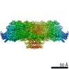
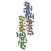
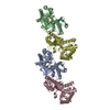
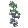
| Title | Crystal structure of phosphoribosylpyrophosphate synthetase from Mycobacterium smegmatis | ||||||
 Components Components | Ribose-phosphate pyrophosphokinase | ||||||
 Keywords Keywords |  TRANSFERASE / phosphoribosylpyrophosphate synthetase / PRPP synthetase / Mycobacterium smegmatis TRANSFERASE / phosphoribosylpyrophosphate synthetase / PRPP synthetase / Mycobacterium smegmatis | ||||||
| Function / homology |  Function and homology informationribose-phosphate diphosphokinase / ribose phosphate diphosphokinase activity / ribonucleoside monophosphate biosynthetic process / nucleotide biosynthetic process / 5-phosphoribose 1-diphosphate biosynthetic process / nucleoside metabolic process / kinase activity / magnesium ion binding / ATP binding / cytoplasm Function and homology informationribose-phosphate diphosphokinase / ribose phosphate diphosphokinase activity / ribonucleoside monophosphate biosynthetic process / nucleotide biosynthetic process / 5-phosphoribose 1-diphosphate biosynthetic process / nucleoside metabolic process / kinase activity / magnesium ion binding / ATP binding / cytoplasmSimilarity search - Function | ||||||
| Biological species |  Mycobacterium smegmatis (bacteria) Mycobacterium smegmatis (bacteria) | ||||||
| Method | X-RAY DIFFRACTION / SYNCHROTRON / MOLECULAR REPLACEMENT / Resolution: 2.4 Å | ||||||
 Authors Authors | Donini, S. / Garavaglia, S. / Ferraris, D.M. / Miggiano, R. / Mori, S. / Shibayama, K. / Rizzi, M. | ||||||
| Funding support |  Italy, 1items Italy, 1items
| ||||||
 Citation Citation | Journal: PLoS ONE / Year: 2017 Title: Biochemical and structural investigations on phosphoribosylpyrophosphate synthetase from Mycobacterium smegmatis. Authors: Donini, S. / Garavaglia, S. / Ferraris, D.M. / Miggiano, R. / Mori, S. / Shibayama, K. / Rizzi, M. | ||||||
| History |
|
- Structure visualization
| Structure viewer | Molecule: MolmilJmol/JSmol |
|---|
- Downloads & links
-Download
| PDBx/mmCIF format | 5mp7.cif.gz | 180.8 KB | Display | PDBx/mmCIF format |
|---|---|---|---|---|
| PDB format | pdb5mp7.ent.gz | 144.9 KB | Display | PDB format |
| PDBx/mmJSON format | 5mp7.json.gz | Tree view | PDBx/mmJSON format | |
| Others |  Other downloads Other downloads |
-Validation report
| Arichive directory | https://data.pdbj.org/pub/pdb/validation_reports/mp/5mp7ftp://data.pdbj.org/pub/pdb/validation_reports/mp/5mp7 | HTTPS FTP |
|---|
-Related structure data
| Related structure data | 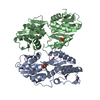 1dkrS S: Starting model for refinement |
|---|---|
| Similar structure data |
-Links
 PDBj
PDBj
- Assembly
| Deposited unit | 
| |||||||||||||||||||||||||||||||||||||||||||||||||||||
|---|---|---|---|---|---|---|---|---|---|---|---|---|---|---|---|---|---|---|---|---|---|---|---|---|---|---|---|---|---|---|---|---|---|---|---|---|---|---|---|---|---|---|---|---|---|---|---|---|---|---|---|---|---|---|
| 1 | 
| |||||||||||||||||||||||||||||||||||||||||||||||||||||
| Unit cell |
| |||||||||||||||||||||||||||||||||||||||||||||||||||||
| Noncrystallographic symmetry (NCS) | NCS domain:
NCS domain segments: Component-ID: 0 / Beg auth comp-ID: LYS / Beg label comp-ID: LYS / End auth comp-ID: GLY / End label comp-ID: GLY / Refine code: 0 / Auth seq-ID: 10 - 316 / Label seq-ID: 10 - 316
NCS ensembles :
|
-Components
| #1: Protein | Mass: 35603.562 Da / Num. of mol.: 3 Source method: isolated from a genetically manipulated source Source: (gene. exp.) Mycobacterium smegmatis (strain ATCC 700084 / mc(2)155) (bacteria)Gene: prs, MSMEG_5427, MSMEI_5278 / Production host: Escherichia coli BL21(DE3) (bacteria)References: UniProt: A0R3C8, ribose-phosphate diphosphokinase#2: Chemical | Acetate  Mass: 59.044 Da / Num. of mol.: 3 / Source method: obtained synthetically / Formula: C2H3O2 Mass: 59.044 Da / Num. of mol.: 3 / Source method: obtained synthetically / Formula: C2H3O2#3: Water | ChemComp-HOH / | Water Mass: 18.015 Da / Num. of mol.: 68 / Source method: isolated from a natural source / Formula: H2O Mass: 18.015 Da / Num. of mol.: 68 / Source method: isolated from a natural source / Formula: H2O |
|---|
-Experimental details
-Experiment
| Experiment | Method: X-RAY DIFFRACTION / Number of used crystals: 1 |
|---|
- Sample preparation
| Crystal | Density Matthews: 2.33 Å3/Da / Density % sol: 47.26 % |
|---|---|
| Crystal grow | Temperature: 277.15 K / Method: vapor diffusion, sitting drop / pH: 4.6 Details: 0.2M NaCl, 0.1M Sodium acetate, 30% (v/v) 2-Methyl-2,4-pentanediol |
-Data collection
| Diffraction | Mean temperature: 100 K |
|---|---|
| Diffraction source | Source: SYNCHROTRON / Site: ESRF  / Beamline: ID23-1 / Wavelength: 0.98 Å / Beamline: ID23-1 / Wavelength: 0.98 Å |
| Detector | Type: DECTRIS PILATUS 6M / Detector: PIXEL / Date: Sep 14, 2013 |
| Radiation | Protocol: SINGLE WAVELENGTH / Monochromatic (M) / Laue (L): M / Scattering type: x-ray |
| Radiation wavelength | Wavelength: 0.98 Å / Relative weight: 1 |
| Reflection | Resolution: 2.4→43.38 Å / Num. obs: 35723 / % possible obs: 96.18 % / Redundancy: 1.9 % / Biso Wilson estimate: 38.07 Å2 / Rmerge(I) obs: 0.067 / Net I/σ(I): 6.24 |
| Reflection shell | Resolution: 2.4→2.49 Å / Redundancy: 1.9 % / Rmerge(I) obs: 0.34 / Mean I/σ(I) obs: 2.34 / % possible all: 97.26 |
- Processing
| Software |
| ||||||||||||||||||||||||||||||||||||||||||||||||||||||||||||||||||||||||||||||||||||||||||||||||||||||||||||||||||||||||||||||||||||||||||||||||||||||||||||||||||||||||||||||||||||||
|---|---|---|---|---|---|---|---|---|---|---|---|---|---|---|---|---|---|---|---|---|---|---|---|---|---|---|---|---|---|---|---|---|---|---|---|---|---|---|---|---|---|---|---|---|---|---|---|---|---|---|---|---|---|---|---|---|---|---|---|---|---|---|---|---|---|---|---|---|---|---|---|---|---|---|---|---|---|---|---|---|---|---|---|---|---|---|---|---|---|---|---|---|---|---|---|---|---|---|---|---|---|---|---|---|---|---|---|---|---|---|---|---|---|---|---|---|---|---|---|---|---|---|---|---|---|---|---|---|---|---|---|---|---|---|---|---|---|---|---|---|---|---|---|---|---|---|---|---|---|---|---|---|---|---|---|---|---|---|---|---|---|---|---|---|---|---|---|---|---|---|---|---|---|---|---|---|---|---|---|---|---|---|---|
| Refinement | Method to determine structure: MOLECULAR REPLACEMENT Starting model: 1DKR Resolution: 2.4→50.01 Å / Cor.coef. Fo:Fc: 0.943 / Cor.coef. Fo:Fc free: 0.93 / SU B: 9.942 / SU ML: 0.22 / Cross valid method: THROUGHOUT / ESU R: 0.524 / ESU R Free: 0.253 / Stereochemistry target values: MAXIMUM LIKELIHOOD / Details: HYDROGENS HAVE BEEN ADDED IN THE RIDING POSITIONS
| ||||||||||||||||||||||||||||||||||||||||||||||||||||||||||||||||||||||||||||||||||||||||||||||||||||||||||||||||||||||||||||||||||||||||||||||||||||||||||||||||||||||||||||||||||||||
| Solvent computation | Ion probe radii: 0.8 Å / Shrinkage radii: 0.8 Å / VDW probe radii: 1.2 Å / Solvent model: MASK | ||||||||||||||||||||||||||||||||||||||||||||||||||||||||||||||||||||||||||||||||||||||||||||||||||||||||||||||||||||||||||||||||||||||||||||||||||||||||||||||||||||||||||||||||||||||
| Displacement parameters | Biso mean: 42.983 Å2
| ||||||||||||||||||||||||||||||||||||||||||||||||||||||||||||||||||||||||||||||||||||||||||||||||||||||||||||||||||||||||||||||||||||||||||||||||||||||||||||||||||||||||||||||||||||||
| Refinement step | Cycle: 1 / Resolution: 2.4→50.01 Å
| ||||||||||||||||||||||||||||||||||||||||||||||||||||||||||||||||||||||||||||||||||||||||||||||||||||||||||||||||||||||||||||||||||||||||||||||||||||||||||||||||||||||||||||||||||||||
| Refine LS restraints |
|