 Movie
Movie Controller

[English] 日本語
 Yorodumi
- PDB-1dkr: CRYSTAL STRUCTURES OF BACILLUS SUBTILIS PHOSPHORIBOSYLPYROPHOSPHA... -
+ Open data

- Basic information
| Entry | Database: PDB / ID: 1dkr | ||||||
|---|---|---|---|---|---|---|---|
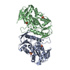
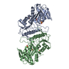
| Title | CRYSTAL STRUCTURES OF BACILLUS SUBTILIS PHOSPHORIBOSYLPYROPHOSPHATE SYNTHETASE: MOLECULAR BASIS OF ALLOSTERIC INHIBITION AND ACTIVATION. | ||||||
 Components Components | PHOSPHORIBOSYL PYROPHOSPHATE SYNTHETASE | ||||||
 Keywords Keywords | TRANSFERASE / DOMAIN DUPLICATION / OPEN ALPHA BETA DOMAIN STRUCTURE / PHOSPHORIBOSYLTRANSFERASE TYPE I FOLD. | ||||||
| Function / homology |  Function and homology information Function and homology informationribose phosphate diphosphokinase complex / ribonucleoside monophosphate biosynthetic process / ribose-phosphate diphosphokinase / ribose phosphate diphosphokinase activity / 5-phosphoribose 1-diphosphate biosynthetic process / purine nucleotide biosynthetic process / kinase activity / magnesium ion binding / ATP binding / cytoplasm Similarity search - Function | ||||||
| Biological species |  | ||||||
| Method |  X-RAY DIFFRACTION / Resolution: 2.3 Å X-RAY DIFFRACTION / Resolution: 2.3 Å | ||||||
 Authors Authors | Eriksen, T.A. / Kadziola, A. / Bentsen, A.-K. / Harlow, K.W. / Larsen, S. | ||||||
 Citation Citation | Journal: Nat.Struct.Biol. / Year: 2000 Title: Structural basis for the function of Bacillus subtilis phosphoribosyl-pyrophosphate synthetase. Authors: Eriksen, T.A. / Kadziola, A. / Bentsen, A.K. / Harlow, K.W. / Larsen, S. #1: Journal: Proteins / Year: 1996Title: Overexpression of the B. Subtilis Phosphoribosylpyrophosphate Synthetase and Crystallization of the Free Enzyme and its Substrate-Effector Complexes Authors: Bentsen, A.-K. / Larsen, T.A. / Kadziola, A. / Larsen, S. / Harlow, K.W. | ||||||
| History |
|
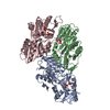
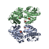
- Structure visualization
| Structure viewer | Molecule: MolmilJmol/JSmol |
|---|
- Downloads & links
-Download
| PDBx/mmCIF format | 1dkr.cif.gz | 129.3 KB | Display | PDBx/mmCIF format |
|---|---|---|---|---|
| PDB format | pdb1dkr.ent.gz | 102.3 KB | Display | PDB format |
| PDBx/mmJSON format | 1dkr.json.gz | Tree view | PDBx/mmJSON format | |
| Others |  Other downloads Other downloads |
-Validation report
| Arichive directory | https://data.pdbj.org/pub/pdb/validation_reports/dk/1dkrftp://data.pdbj.org/pub/pdb/validation_reports/dk/1dkr | HTTPS FTP |
|---|
-Related structure data


-Links
 PDBj
PDBj
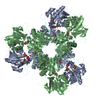
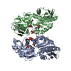
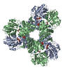
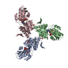
- Assembly
| Deposited unit | 
| ||||||||
|---|---|---|---|---|---|---|---|---|---|
| 1 | 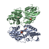
| ||||||||
| Unit cell |
|
-Components
| #1: Protein | Mass: 34910.223 Da / Num. of mol.: 2 Source method: isolated from a genetically manipulated source Source: (gene. exp.) References: UniProt: P14193, ribose-phosphate diphosphokinase #2: Chemical | ChemComp-SO4 /   Mass: 96.063 Da / Num. of mol.: 4 / Source method: obtained synthetically / Formula: SO4 Mass: 96.063 Da / Num. of mol.: 4 / Source method: obtained synthetically / Formula: SO4#3: Water | ChemComp-HOH / |  Mass: 18.015 Da / Num. of mol.: 230 / Source method: isolated from a natural source / Formula: H2O Mass: 18.015 Da / Num. of mol.: 230 / Source method: isolated from a natural source / Formula: H2O |
|---|
-Experimental details
-Experiment
| Experiment | Method: X-RAY DIFFRACTION / Number of used crystals: 1 |
|---|
- Sample preparation
| Crystal | Density Matthews: 2.98 Å3/Da / Density % sol: 58.67 % | ||||||||||||||||||||||||||||||||||||||||||
|---|---|---|---|---|---|---|---|---|---|---|---|---|---|---|---|---|---|---|---|---|---|---|---|---|---|---|---|---|---|---|---|---|---|---|---|---|---|---|---|---|---|---|---|
| Crystal grow | Temperature: 298 K / Method: vapor diffusion, sitting drop / pH: 7.5 Details: AMMONIUM SULFATE; TRISHCL; PEG 400; SODIUM PHOSPHATE; BETA-OCTYLGLUCOSIDE; ALPHA, BETA-METHYLENE ATP; RIBOSE-5-PHOSPHATE; MAGNESIUM SULFATE; ADP, pH 7.5, VAPOR DIFFUSION, SITTING DROP, temperature 298K | ||||||||||||||||||||||||||||||||||||||||||
| Crystal grow | *PLUS pH: 8 | ||||||||||||||||||||||||||||||||||||||||||
| Components of the solutions | *PLUS
|
-Data collection
| Diffraction | Mean temperature: 274 K |
|---|---|
| Diffraction source | Source: ROTATING ANODE / Type: RIGAKU RU200 / Wavelength: 1.5418 |
| Detector | Type: RIGAKU RAXIS II / Detector: IMAGE PLATE / Date: May 23, 1994 |
| Radiation | Protocol: SINGLE WAVELENGTH / Monochromatic (M) / Laue (L): M / Scattering type: x-ray |
| Radiation wavelength | Wavelength: 1.5418 Å / Relative weight: 1 |
| Reflection | Resolution: 2.2→100 Å / Num. all: 40947 / Num. obs: 40947 / % possible obs: 98.8 % / Observed criterion σ(F): 0 / Observed criterion σ(I): 0 / Redundancy: 3.4 % / Biso Wilson estimate: 22.1 Å2 / Rmerge(I) obs: 0.077 / Net I/σ(I): 9 |
| Reflection shell | Resolution: 2.3→2.42 Å / Redundancy: 3.8 % / Rmerge(I) obs: 0.2 / % possible all: 99.6 |
| Reflection | *PLUS Num. obs: 35812 / Rmerge(I) obs: 0.068 |
| Reflection shell | *PLUS % possible obs: 99.6 % / Num. unique obs: 5098 / Rmerge(I) obs: 0.2 / Mean I/σ(I) obs: 3.6 |
- Processing
| Software |
| ||||||||||||||||||||||||||||||||||||||||||||||||||||||||||||
|---|---|---|---|---|---|---|---|---|---|---|---|---|---|---|---|---|---|---|---|---|---|---|---|---|---|---|---|---|---|---|---|---|---|---|---|---|---|---|---|---|---|---|---|---|---|---|---|---|---|---|---|---|---|---|---|---|---|---|---|---|---|
| Refinement | Resolution: 2.3→30 Å / σ(F): 0 / σ(I): 0 / Stereochemistry target values: ENGH & HUBER
| ||||||||||||||||||||||||||||||||||||||||||||||||||||||||||||
| Refinement step | Cycle: LAST / Resolution: 2.3→30 Å
| ||||||||||||||||||||||||||||||||||||||||||||||||||||||||||||
| Refine LS restraints |
| ||||||||||||||||||||||||||||||||||||||||||||||||||||||||||||
| Software | *PLUS Name: X-PLOR / Version: 3.1 / Classification: refinement | ||||||||||||||||||||||||||||||||||||||||||||||||||||||||||||
| Refinement | *PLUS Rfactor obs: 0.18 / Rfactor Rfree: 0.24 / Rfactor Rwork: 0.18 | ||||||||||||||||||||||||||||||||||||||||||||||||||||||||||||
| Solvent computation | *PLUS | ||||||||||||||||||||||||||||||||||||||||||||||||||||||||||||
| Displacement parameters | *PLUS | ||||||||||||||||||||||||||||||||||||||||||||||||||||||||||||
| Refine LS restraints | *PLUS
|
