 Movie
Movie Controller

[English] 日本語
 Yorodumi
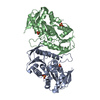
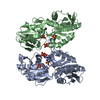
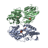
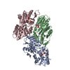
- PDB-3efh: Crystal structure of human phosphoribosyl pyrophosphate synthetase 1 -
+ Open data

- Basic information
| Entry | Database: PDB / ID: 3efh | ||||||
|---|---|---|---|---|---|---|---|
| Title | Crystal structure of human phosphoribosyl pyrophosphate synthetase 1 | ||||||
 Components Components | Ribose-phosphate pyrophosphokinase 1 | ||||||
 Keywords Keywords | TRANSFERASE / Charcot-Marie-Tooth disease / Deafness / Disease mutation / Gout / Kinase / Magnesium / Metal-binding / Nucleotide biosynthesis / Polymorphism | ||||||
| Function / homology |  Function and homology information Function and homology information5-Phosphoribose 1-diphosphate biosynthesis / hypoxanthine biosynthetic process / ribose phosphate diphosphokinase complex / ribonucleoside monophosphate biosynthetic process / ribose-phosphate diphosphokinase / ribose phosphate diphosphokinase activity / urate biosynthetic process / 5-phosphoribose 1-diphosphate biosynthetic process / pyrimidine nucleotide biosynthetic process / purine nucleotide biosynthetic process ...5-Phosphoribose 1-diphosphate biosynthesis / hypoxanthine biosynthetic process / ribose phosphate diphosphokinase complex / ribonucleoside monophosphate biosynthetic process / ribose-phosphate diphosphokinase / ribose phosphate diphosphokinase activity / urate biosynthetic process / 5-phosphoribose 1-diphosphate biosynthetic process / pyrimidine nucleotide biosynthetic process / purine nucleotide biosynthetic process / purine nucleobase metabolic process / kinase activity / nervous system development / magnesium ion binding / protein homodimerization activity / ATP binding / identical protein binding / cytoplasm / cytosol Similarity search - Function | ||||||
| Biological species |  Homo sapiens (human) Homo sapiens (human) | ||||||
| Method |  X-RAY DIFFRACTION / MOLECULAR REPLACEMENT / Resolution: 2.6 Å X-RAY DIFFRACTION / MOLECULAR REPLACEMENT / Resolution: 2.6 Å | ||||||
 Authors Authors | Li, X. / Peng, X. / Li, Y. | ||||||
 Citation Citation | Journal: To be Published Title: Crystal structure of human phosphoribosyl pyrophosphate synthetase 1 Authors: Peng, X. / Li, X. | ||||||
| History |
|
- Structure visualization
| Structure viewer | Molecule: MolmilJmol/JSmol |
|---|
- Downloads & links
-Download
| PDBx/mmCIF format | 3efh.cif.gz | 126.7 KB | Display | PDBx/mmCIF format |
|---|---|---|---|---|
| PDB format | pdb3efh.ent.gz | 98.1 KB | Display | PDB format |
| PDBx/mmJSON format | 3efh.json.gz | Tree view | PDBx/mmJSON format | |
| Others |  Other downloads Other downloads |
-Validation report
| Arichive directory | https://data.pdbj.org/pub/pdb/validation_reports/ef/3efhftp://data.pdbj.org/pub/pdb/validation_reports/ef/3efh | HTTPS FTP |
|---|
-Related structure data
| Related structure data |  2hcrS S: Starting model for refinement |
|---|---|
| Similar structure data |

-Links
 PDBj
PDBj
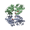
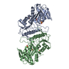
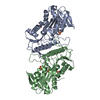
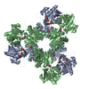
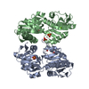
- Assembly
| Deposited unit | 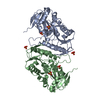
| ||||||||
|---|---|---|---|---|---|---|---|---|---|
| 1 | 
| ||||||||
| 2 | 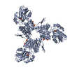
| ||||||||
| 3 | 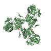
| ||||||||
| Unit cell |
|
-Components
| #1: Protein | Mass: 35950.375 Da / Num. of mol.: 2 Source method: isolated from a genetically manipulated source Source: (gene. exp.) Homo sapiens (human) / Gene: PRPS1 / Plasmid: pET-22b(+) / Production host:  References: UniProt: P60891, ribose-phosphate diphosphokinase #2: Chemical | ChemComp-SO4 /   Mass: 96.063 Da / Num. of mol.: 6 / Source method: obtained synthetically / Formula: SO4 Mass: 96.063 Da / Num. of mol.: 6 / Source method: obtained synthetically / Formula: SO4#3: Water | ChemComp-HOH / |  Mass: 18.015 Da / Num. of mol.: 108 / Source method: isolated from a natural source / Formula: H2O Mass: 18.015 Da / Num. of mol.: 108 / Source method: isolated from a natural source / Formula: H2O |
|---|
-Experimental details
-Experiment
| Experiment | Method: X-RAY DIFFRACTION / Number of used crystals: 1 |
|---|
- Sample preparation
| Crystal | Density Matthews: 2.44 Å3/Da / Density % sol: 49.62 % |
|---|---|
| Crystal grow | Temperature: 295 K / Method: vapor diffusion, hanging drop / pH: 4.3 Details: 2.1M ammonium sulfate, 0.1M sodium acetate, 10% (v/v) glycerol, pH 4.3, VAPOR DIFFUSION, HANGING DROP, temperature 295K |
-Data collection
| Diffraction | Mean temperature: 100 K |
|---|---|
| Diffraction source | Source: ROTATING ANODE / Type: RIGAKU MICROMAX-007 / Wavelength: 1.54179 Å |
| Detector | Type: MAR scanner 345 mm plate / Detector: IMAGE PLATE / Date: Nov 18, 2005 |
| Radiation | Protocol: SINGLE WAVELENGTH / Monochromatic (M) / Laue (L): M / Scattering type: x-ray |
| Radiation wavelength | Wavelength: 1.54179 Å / Relative weight: 1 |
| Reflection | Resolution: 2.6→42.295 Å / Num. all: 18873 / Num. obs: 18518 / % possible obs: 91.2 % / Observed criterion σ(F): 0 / Observed criterion σ(I): 0 / Redundancy: 1.4 % / Biso Wilson estimate: 50.4 Å2 / Rmerge(I) obs: 0.065 / Rsym value: 0.065 / Net I/σ(I): 8.5 |
| Reflection shell | Resolution: 2.6→2.74 Å / Redundancy: 1.4 % / Rmerge(I) obs: 0.345 / Mean I/σ(I) obs: 1.8 / Num. unique all: 2623 / Rsym value: 0.345 / % possible all: 88.5 |
- Processing
| Software |
| ||||||||||||||||||||||||||||||||||||||||||||||||||||||||||||||||||||||||||||||||||||||||||||||||||||||||||||||||||||||||||||||||||||||||||||||||||||||||||||||||||||||||||
|---|---|---|---|---|---|---|---|---|---|---|---|---|---|---|---|---|---|---|---|---|---|---|---|---|---|---|---|---|---|---|---|---|---|---|---|---|---|---|---|---|---|---|---|---|---|---|---|---|---|---|---|---|---|---|---|---|---|---|---|---|---|---|---|---|---|---|---|---|---|---|---|---|---|---|---|---|---|---|---|---|---|---|---|---|---|---|---|---|---|---|---|---|---|---|---|---|---|---|---|---|---|---|---|---|---|---|---|---|---|---|---|---|---|---|---|---|---|---|---|---|---|---|---|---|---|---|---|---|---|---|---|---|---|---|---|---|---|---|---|---|---|---|---|---|---|---|---|---|---|---|---|---|---|---|---|---|---|---|---|---|---|---|---|---|---|---|---|---|---|---|---|
| Refinement | Method to determine structure: MOLECULAR REPLACEMENT Starting model: PDB ENTRY 2HCR Resolution: 2.6→30 Å / Cor.coef. Fo:Fc: 0.916 / Cor.coef. Fo:Fc free: 0.91 / SU B: 8.566 / SU ML: 0.184 / Cross valid method: THROUGHOUT / σ(F): 0 / σ(I): 0 / ESU R Free: 0.328 / Stereochemistry target values: MAXIMUM LIKELIHOOD / Details: HYDROGENS HAVE BEEN ADDED IN THE RIDING POSITIONS
| ||||||||||||||||||||||||||||||||||||||||||||||||||||||||||||||||||||||||||||||||||||||||||||||||||||||||||||||||||||||||||||||||||||||||||||||||||||||||||||||||||||||||||
| Solvent computation | Ion probe radii: 0.8 Å / Shrinkage radii: 0.8 Å / VDW probe radii: 1.4 Å / Solvent model: MASK | ||||||||||||||||||||||||||||||||||||||||||||||||||||||||||||||||||||||||||||||||||||||||||||||||||||||||||||||||||||||||||||||||||||||||||||||||||||||||||||||||||||||||||
| Displacement parameters | Biso mean: 33.116 Å2
| ||||||||||||||||||||||||||||||||||||||||||||||||||||||||||||||||||||||||||||||||||||||||||||||||||||||||||||||||||||||||||||||||||||||||||||||||||||||||||||||||||||||||||
| Refinement step | Cycle: LAST / Resolution: 2.6→30 Å
| ||||||||||||||||||||||||||||||||||||||||||||||||||||||||||||||||||||||||||||||||||||||||||||||||||||||||||||||||||||||||||||||||||||||||||||||||||||||||||||||||||||||||||
| Refine LS restraints |
| ||||||||||||||||||||||||||||||||||||||||||||||||||||||||||||||||||||||||||||||||||||||||||||||||||||||||||||||||||||||||||||||||||||||||||||||||||||||||||||||||||||||||||
| LS refinement shell | Resolution: 2.6→2.667 Å / Total num. of bins used: 20
|
