 Movie
Movie Controller

[English] 日本語
 Yorodumi
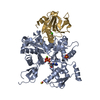
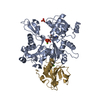
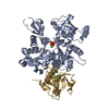
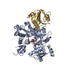
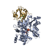
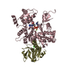
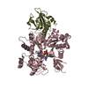
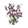
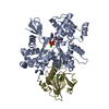
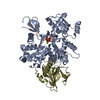
- PDB-1yvn: THE YEAST ACTIN VAL 159 ASN MUTANT COMPLEX WITH HUMAN GELSOLIN SE... -
+ Open data

- Basic information
| Entry | Database: PDB / ID: 1yvn | ||||||
|---|---|---|---|---|---|---|---|
| Title | THE YEAST ACTIN VAL 159 ASN MUTANT COMPLEX WITH HUMAN GELSOLIN SEGMENT 1. | ||||||
 Components Components |
| ||||||
 Keywords Keywords |  STRUCTURAL PROTEIN / YEAST / ACTIN / MUTANT V159N / GELSOLIN / ACTIN-BINDING / MG-ATP STRUCTURAL PROTEIN / YEAST / ACTIN / MUTANT V159N / GELSOLIN / ACTIN-BINDING / MG-ATP | ||||||
| Function / homology |  Function and homology information Function and homology informationmitotic actomyosin contractile ring contraction / RHOA GTPase cycle / cellular bud neck contractile ring / RHO GTPases activate IQGAPs / RHO GTPases Activate WASPs and WAVEs / Regulation of actin dynamics for phagocytic cup formation / ascospore wall assembly / vacuole inheritance / striated muscle atrophy / regulation of establishment of T cell polarity ...mitotic actomyosin contractile ring contraction / RHOA GTPase cycle / cellular bud neck contractile ring / RHO GTPases activate IQGAPs / RHO GTPases Activate WASPs and WAVEs / Regulation of actin dynamics for phagocytic cup formation / ascospore wall assembly / vacuole inheritance / striated muscle atrophy / regulation of establishment of T cell polarity / regulation of plasma membrane raft polarization / regulation of receptor clustering / renal protein absorption / positive regulation of keratinocyte apoptotic process / positive regulation of protein processing in phagocytic vesicle / positive regulation of actin nucleation / phosphatidylinositol 3-kinase catalytic subunit binding / positive regulation of cysteine-type endopeptidase activity involved in apoptotic signaling pathway / actin cap / actin cortical patch / Swr1 complex / sequestering of actin monomers / regulation of podosome assembly / myosin II binding / Platelet degranulation / : / negative regulation of viral entry into host cell / actin filament severing / Ino80 complex / actin filament capping / barbed-end actin filament capping / actin filament depolymerization / actin polymerization or depolymerization / cell projection assembly / cardiac muscle cell contraction / podosome / Sensory processing of sound by outer hair cells of the cochlea / NuA4 histone acetyltransferase complex / relaxation of cardiac muscle / phagocytosis, engulfment / cortical actin cytoskeleton / establishment of cell polarity / hepatocyte apoptotic process / actin filament bundle / cilium assembly / protein secretion / sarcoplasm / Caspase-mediated cleavage of cytoskeletal proteins / phagocytic vesicle / phosphatidylinositol-4,5-bisphosphate binding / response to muscle stretch / actin filament polymerization / central nervous system development / actin filament organization / actin filament / Hydrolases; Acting on acid anhydrides; Acting on acid anhydrides to facilitate cellular and subcellular movement / protein destabilization / structural constituent of cytoskeleton / cellular response to type II interferon / endocytosis / actin filament binding / actin cytoskeleton / lamellipodium / actin binding / blood microparticle / secretory granule lumen / ficolin-1-rich granule lumen / amyloid fibril formation / hydrolase activity / chromatin remodeling / Amyloid fiber formation / focal adhesion / DNA repair / DNA-templated transcription / calcium ion binding / chromatin / Neutrophil degranulation / positive regulation of gene expression / regulation of DNA-templated transcription / extracellular space / extracellular exosome / extracellular region / ATP binding / identical protein binding / nucleus / plasma membrane / cytosol / cytoplasmSimilarity search - Function | ||||||
| Biological species |  Homo sapiens (human) Homo sapiens (human) Saccharomyces cerevisiae (brewer's yeast) Saccharomyces cerevisiae (brewer's yeast) | ||||||
| Method | X-RAY DIFFRACTION / SYNCHROTRON / MOLECULAR REPLACEMENT / Resolution: 2.1 Å | ||||||
 Authors Authors | Vorobiev, S.M. / Belmont, L.D. / Drubin, D.G. / Almo, S.C. | ||||||
 Citation Citation | Journal: To be Published Title: Crystal structure of yeast actin V159N mutant Authors: Vorobiev, S.M. / Belmont, L.D. / Drubin, D.D. / Almos, C. #1: Journal: Proc.Natl.Acad.Sci.USA / Year: 1999 Title: A change in actin conformation associated with filament instability after Pi release. Authors: Belmont, L.D. / Orlova, A. / Drubin, D.G. / Egelman, E.H. #2: Journal: Nature / Year: 1993Title: Structure of gelsolin segment 1-actin complex and the mechanism of filament severing. Authors: McLaughlin, P.J. / Gooch, J.T. / Mannherz, H.G. / Weeds, A.G. #3: Journal: Nature / Year: 1990Title: Atomic structure of the actin:DNase I complex. Authors: Kabsch, W. / Mannherz, H.G. / Suck, D. / Pai, E.F. / Holmes, K.C. | ||||||
| History |
|
- Structure visualization
| Structure viewer | Molecule: MolmilJmol/JSmol |
|---|
- Downloads & links
-Download
| PDBx/mmCIF format | 1yvn.cif.gz | 119.8 KB | Display | PDBx/mmCIF format |
|---|---|---|---|---|
| PDB format | pdb1yvn.ent.gz | 89.7 KB | Display | PDB format |
| PDBx/mmJSON format | 1yvn.json.gz | Tree view | PDBx/mmJSON format | |
| Others |  Other downloads Other downloads |
-Validation report
| Arichive directory | https://data.pdbj.org/pub/pdb/validation_reports/yv/1yvnftp://data.pdbj.org/pub/pdb/validation_reports/yv/1yvn | HTTPS FTP |
|---|
-Related structure data
| Related structure data |  1yagS S: Starting model for refinement |
|---|---|
| Similar structure data |
-Links
 PDBj
PDBj


- Assembly
| Deposited unit | 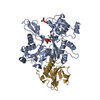
| ||||||||
|---|---|---|---|---|---|---|---|---|---|
| 1 |
| ||||||||
| 2 | 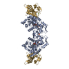
| ||||||||
| Unit cell |
| ||||||||
| Components on special symmetry positions |
|
-Components
-Protein , 2 types, 2 molecules AG
| #1: Protein | Mass: 41750.520 Da / Num. of mol.: 1 / Mutation: V159N / Source method: isolated from a natural source / Details: THE GENE NAME IS ACT. / Source: (natural) Saccharomyces cerevisiae (brewer's yeast) / Strain: ACT1-159 / References: UniProt: P60010 |
|---|---|
| #2: Protein | Mass: 14071.831 Da / Num. of mol.: 1 / Fragment: FRAGMENT 1 Source method: isolated from a genetically manipulated source Details: CA(2+)-BOUND PROTEIN / Source: (gene. exp.) Homo sapiens (human) / Description: SYNTHETIC GENE / Plasmid: PMW172 / Species (production host): Escherichia coli / Production host:  Escherichia coli BL21(DE3) (bacteria) / Strain (production host): BL21(DE3) / References: UniProt: P06396 Escherichia coli BL21(DE3) (bacteria) / Strain (production host): BL21(DE3) / References: UniProt: P06396 |
-Non-polymers , 5 types, 287 molecules 


| #3: Chemical | ChemComp-MG /  Mass: 24.305 Da / Num. of mol.: 1 / Source method: obtained synthetically / Formula: Mg Mass: 24.305 Da / Num. of mol.: 1 / Source method: obtained synthetically / Formula: Mg |
|---|---|
| #4: Chemical | ChemComp-SO4 / Sulfate Mass: 96.063 Da / Num. of mol.: 1 / Source method: obtained synthetically / Formula: SO4 Mass: 96.063 Da / Num. of mol.: 1 / Source method: obtained synthetically / Formula: SO4 |
| #5: Chemical | ChemComp-ATP / Adenosine triphosphate Mass: 507.181 Da / Num. of mol.: 1 / Source method: obtained synthetically / Formula: C10H16N5O13P3 / Comment: ATP, energy-carrying molecule*YM Mass: 507.181 Da / Num. of mol.: 1 / Source method: obtained synthetically / Formula: C10H16N5O13P3 / Comment: ATP, energy-carrying molecule*YM |
| #6: Chemical | ChemComp-CA /  Mass: 40.078 Da / Num. of mol.: 1 / Source method: obtained synthetically / Formula: Ca Mass: 40.078 Da / Num. of mol.: 1 / Source method: obtained synthetically / Formula: Ca |
| #7: Water | ChemComp-HOH / WaterMass: 18.015 Da / Num. of mol.: 283 / Source method: isolated from a natural source / Formula: H2O |
-Experimental details
-Experiment
| Experiment | Method: X-RAY DIFFRACTION / Number of used crystals: 1 |
|---|
- Sample preparation
| Crystal | Density Matthews: 2.51 Å3/Da / Density % sol: 50.55 % |
|---|---|
| Crystal grow | pH: 8 Details: 0.1 M HEPES, 1.60 M (NH4)2SO4, 2 MM MG-ATP, PH=8.0. |
-Data collection
| Diffraction | Mean temperature: 105 K |
|---|---|
| Diffraction source | Source: SYNCHROTRON / Site: NSLS  / Beamline: X9B / Wavelength: 1.0702 / Beamline: X9B / Wavelength: 1.0702 |
| Detector | Type: ADSC QUANTUM 4 / Detector: CCD / Date: Feb 1, 1999 |
| Radiation | Protocol: SINGLE WAVELENGTH / Monochromatic (M) / Laue (L): M / Scattering type: x-ray |
| Radiation wavelength | Wavelength: 1.0702 Å / Relative weight: 1 |
| Reflection | Resolution: 2.1→25 Å / Num. obs: 33130 / % possible obs: 86.8 % / Redundancy: 2.95 % / Rmerge(I) obs: 0.057 |
- Processing
| Software |
| ||||||||||||||||||||||||||||||||||||||||||||||||||||||||||||
|---|---|---|---|---|---|---|---|---|---|---|---|---|---|---|---|---|---|---|---|---|---|---|---|---|---|---|---|---|---|---|---|---|---|---|---|---|---|---|---|---|---|---|---|---|---|---|---|---|---|---|---|---|---|---|---|---|---|---|---|---|---|
| Refinement | Method to determine structure: MOLECULAR REPLACEMENT Starting model: PDB ENTRY 1YAG Resolution: 2.1→25 Å / Cross valid method: THROUGHOUT / σ(F): 0
| ||||||||||||||||||||||||||||||||||||||||||||||||||||||||||||
| Displacement parameters | Biso mean: 22.69 Å2 | ||||||||||||||||||||||||||||||||||||||||||||||||||||||||||||
| Refinement step | Cycle: LAST / Resolution: 2.1→25 Å
| ||||||||||||||||||||||||||||||||||||||||||||||||||||||||||||
| Refine LS restraints |
| ||||||||||||||||||||||||||||||||||||||||||||||||||||||||||||
| LS refinement shell | Resolution: 2.1→2.2 Å / Total num. of bins used: 8
| ||||||||||||||||||||||||||||||||||||||||||||||||||||||||||||
| Xplor file |
|
