 Movie
Movie Controller

[English] 日本語
 Yorodumi
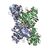
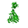
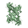
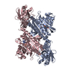
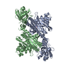
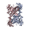
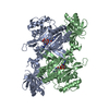
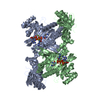
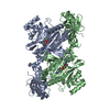
- PDB-1e1o: lysyl-tRNA Synthetase (LYSU) hexagonal form, complexed with lysine -
+ Open data

- Basic information
| Entry | Database: PDB / ID: 1e1o | ||||||
|---|---|---|---|---|---|---|---|
| Title | lysyl-tRNA Synthetase (LYSU) hexagonal form, complexed with lysine | ||||||
 Components Components | LYSYL-TRNA SYNTHETASE, HEAT INDUCIBLE | ||||||
 Keywords Keywords |  LIGASE / AMINOACYL-TRNA SYNTHETASE / PROTEIN BIOSYNTHESIS LIGASE / AMINOACYL-TRNA SYNTHETASE / PROTEIN BIOSYNTHESIS | ||||||
| Function / homology |  Function and homology informationRNA capping / lysine-tRNA ligase / lysine-tRNA ligase activity / lysyl-tRNA aminoacylation / tRNA aminoacylation for protein translation / ligase activity / cellular response to heat / tRNA binding / magnesium ion binding / protein homodimerization activity ...RNA capping / lysine-tRNA ligase / lysine-tRNA ligase activity / lysyl-tRNA aminoacylation / tRNA aminoacylation for protein translation / ligase activity / cellular response to heat / tRNA binding / magnesium ion binding / protein homodimerization activity / ATP binding / membrane / cytosol Function and homology informationRNA capping / lysine-tRNA ligase / lysine-tRNA ligase activity / lysyl-tRNA aminoacylation / tRNA aminoacylation for protein translation / ligase activity / cellular response to heat / tRNA binding / magnesium ion binding / protein homodimerization activity ...RNA capping / lysine-tRNA ligase / lysine-tRNA ligase activity / lysyl-tRNA aminoacylation / tRNA aminoacylation for protein translation / ligase activity / cellular response to heat / tRNA binding / magnesium ion binding / protein homodimerization activity / ATP binding / membrane / cytosolSimilarity search - Function | ||||||
| Biological species |  ESCHERICHIA COLI (E. coli) ESCHERICHIA COLI (E. coli) | ||||||
| Method | X-RAY DIFFRACTION / SYNCHROTRON / MOLECULAR REPLACEMENT / Resolution: 2.12 Å | ||||||
 Authors Authors | Desogus, G. / Todone, F. / Brick, P. / Onesti, S. | ||||||
 Citation Citation | Journal: Biochemistry / Year: 2000 Title: Active Site of Lysyl-tRNA Synthetase: Structural Studies of the Adenylation Reaction Authors: Desogus, G. / Todone, F. / Brick, P. / Onesti, S. | ||||||
| History |
|
- Structure visualization
| Structure viewer | Molecule: MolmilJmol/JSmol |
|---|
- Downloads & links
-Download
| PDBx/mmCIF format | 1e1o.cif.gz | 118 KB | Display | PDBx/mmCIF format |
|---|---|---|---|---|
| PDB format | pdb1e1o.ent.gz | 90.6 KB | Display | PDB format |
| PDBx/mmJSON format | 1e1o.json.gz | Tree view | PDBx/mmJSON format | |
| Others |  Other downloads Other downloads |
-Validation report
| Arichive directory | https://data.pdbj.org/pub/pdb/validation_reports/e1/1e1oftp://data.pdbj.org/pub/pdb/validation_reports/e1/1e1o | HTTPS FTP |
|---|
-Related structure data
| Related structure data | 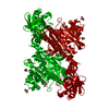 1e1tC  1e22C 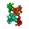 1e24C  1lylS S: Starting model for refinement C: citing same article ( |
|---|---|
| Similar structure data |

-Links
 PDBj
PDBj

- Assembly
| Deposited unit | 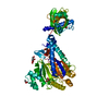
| ||||||||
|---|---|---|---|---|---|---|---|---|---|
| 1 | 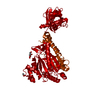
| ||||||||
| Unit cell |
| ||||||||
| Details | THE PROTEIN IS AN ACTIVE DIMER. IN THE HEXAGONAL CELL THEMOLECULAR DYADE COINCIDE WITH A CRYSTALLOGRAPHIC 2-FOLDAXIS. TO GENERATE THE BIOLOGICALLY FUNCTIONAL DIMER THESYMMETRY OPERATION Y , X, -Z+1/3 NEEDS TO BE APPLIED TO THEATOMIC COORDINATES. |
-Components
| #1: Protein | Mass: 57767.191 Da / Num. of mol.: 1 Source method: isolated from a genetically manipulated source Source: (gene. exp.) ESCHERICHIA COLI (E. coli) / Strain: K-12 / Cellular location: CYTOPLASM / Gene: LYSU / Plasmid: PXLYS5 / Gene (production host): LYSU / Production host: ESCHERICHIA COLI (E. coli) / Strain (production host): TG2 / References: UniProt: P0A8N5, lysine-tRNA ligase | ||||
|---|---|---|---|---|---|
| #2: Chemical | ChemComp-LYS / Lysine  Type: L-peptide linking / Mass: 147.195 Da / Num. of mol.: 1 / Source method: obtained synthetically / Formula: C6H15N2O2 Type: L-peptide linking / Mass: 147.195 Da / Num. of mol.: 1 / Source method: obtained synthetically / Formula: C6H15N2O2 | ||||
| #3: Chemical | ChemComp-GOL / Glycerol  Mass: 92.094 Da / Num. of mol.: 8 / Source method: obtained synthetically / Formula: C3H8O3 Mass: 92.094 Da / Num. of mol.: 8 / Source method: obtained synthetically / Formula: C3H8O3#4: Water | ChemComp-HOH / | Water Mass: 18.015 Da / Num. of mol.: 361 / Source method: isolated from a natural source / Formula: H2O Mass: 18.015 Da / Num. of mol.: 361 / Source method: isolated from a natural source / Formula: H2OCompound details | THE STRUCTURE OF E. COLI LYSYL-TRNA SYNTHETASE LYSU BOUND TO THE SUBSTRATE LYSINE HAS BEEN SOLVED ...THE STRUCTURE OF E. COLI LYSYL-TRNA SYNTHETASE | |
-Experimental details
-Experiment
| Experiment | Method: X-RAY DIFFRACTION / Number of used crystals: 1 |
|---|
- Sample preparation
| Crystal | Density Matthews: 4.8 Å3/Da / Density % sol: 74 % Description: STRUCTURE WAS SOLVED BY USING 1LYL COORDINATED TRANSLATED TO ACCOUNT FOR THE DIFFERENT ORIGIN OF THE RELATED HEXAGONAL CELL | ||||||||||||||||||||||||||||||||||||||||||||||||
|---|---|---|---|---|---|---|---|---|---|---|---|---|---|---|---|---|---|---|---|---|---|---|---|---|---|---|---|---|---|---|---|---|---|---|---|---|---|---|---|---|---|---|---|---|---|---|---|---|---|
| Crystal grow | pH: 6.8 Details: PROTEIN WAS CRYSTALLISED FROM 0.1M PIPES PH 6.8, 0.5 M LICL; 20% PEG 4K, 17% GLYCEROL | ||||||||||||||||||||||||||||||||||||||||||||||||
| Crystal grow | *PLUS pH: 7.5 / Method: vapor diffusion, hanging drop | ||||||||||||||||||||||||||||||||||||||||||||||||
| Components of the solutions | *PLUS
|
-Data collection
| Diffraction | Mean temperature: 100 K |
|---|---|
| Diffraction source | Source: SYNCHROTRON / Site: EMBL/DESY, HAMBURG  / Beamline: BW7B / Wavelength: 0.86 / Beamline: BW7B / Wavelength: 0.86 |
| Detector | Type: MARRESEARCH / Detector: IMAGE PLATE / Date: Jun 15, 1995 / Details: MIRROR |
| Radiation | Monochromator: SI(111) / Protocol: SINGLE WAVELENGTH / Monochromatic (M) / Laue (L): M / Scattering type: x-ray |
| Radiation wavelength | Wavelength: 0.86 Å / Relative weight: 1 |
| Reflection | Resolution: 2.12→25 Å / Num. obs: 60434 / % possible obs: 99.6 % / Redundancy: 16.2 % / Rmerge(I) obs: 0.085 / Rsym value: 0.085 / Net I/σ(I): 8.8 |
| Reflection shell | Resolution: 2.12→2.16 Å / Redundancy: 15.4 % / Rmerge(I) obs: 0.26 / Mean I/σ(I) obs: 3.7 / Rsym value: 0.26 / % possible all: 99.6 |
| Reflection | *PLUS Num. measured all: 977338 |
| Reflection shell | *PLUS % possible obs: 99.6 % |
- Processing
| Software |
| ||||||||||||||||||||||||||||||||||||||||||||||||||||||||||||||||||||||||||||||||
|---|---|---|---|---|---|---|---|---|---|---|---|---|---|---|---|---|---|---|---|---|---|---|---|---|---|---|---|---|---|---|---|---|---|---|---|---|---|---|---|---|---|---|---|---|---|---|---|---|---|---|---|---|---|---|---|---|---|---|---|---|---|---|---|---|---|---|---|---|---|---|---|---|---|---|---|---|---|---|---|---|---|
| Refinement | Method to determine structure: MOLECULAR REPLACEMENT Starting model: PDB ENTRY 1LYL Resolution: 2.12→20 Å / Rfactor Rfree error: 0.005 / Isotropic thermal model: RESTRAINED / Cross valid method: THROUGHOUT / σ(F): 0
| ||||||||||||||||||||||||||||||||||||||||||||||||||||||||||||||||||||||||||||||||
| Refine analyze | Luzzati d res low obs: 20 Å | ||||||||||||||||||||||||||||||||||||||||||||||||||||||||||||||||||||||||||||||||
| Refinement step | Cycle: LAST / Resolution: 2.12→20 Å
| ||||||||||||||||||||||||||||||||||||||||||||||||||||||||||||||||||||||||||||||||
| Refine LS restraints |
| ||||||||||||||||||||||||||||||||||||||||||||||||||||||||||||||||||||||||||||||||
| LS refinement shell | Resolution: 2.12→2.22 Å / Rfactor Rfree error: 0.02 / Total num. of bins used: 8
| ||||||||||||||||||||||||||||||||||||||||||||||||||||||||||||||||||||||||||||||||
| Xplor file |
| ||||||||||||||||||||||||||||||||||||||||||||||||||||||||||||||||||||||||||||||||
| Software | *PLUS Name: X-PLOR / Version: 3.1 / Classification: refinement | ||||||||||||||||||||||||||||||||||||||||||||||||||||||||||||||||||||||||||||||||
| Refine LS restraints | *PLUS
|
