- PDB-1e24: LYSYL-TRNA SYNTHETASE (LYSU) HEXAGONAL FORM complexed with lysine... -
+
Open data
ID or keywords:
Loading...
-
Basic information
Entry
Database: PDB / ID: 1.0E+24
Title
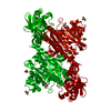
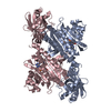
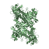
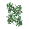
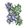
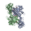
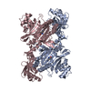
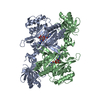
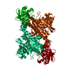
LYSYL-TRNA SYNTHETASE (LYSU) HEXAGONAL FORM complexed with lysine and ATP and MN2+
Components
LYSYL-TRNA SYNTHETASE
Keywords
LIGASE / AMINOACYL-TRNA SYNTHETASE / PROTEIN BIOSYNTHESIS
Function / homology
Function and homology information
RNA capping / lysine-tRNA ligase / lysine-tRNA ligase activity / lysyl-tRNA aminoacylation / tRNA aminoacylation for protein translation / ligase activity / cellular response to heat / nucleic acid binding / tRNA binding / magnesium ion binding ...RNA capping / lysine-tRNA ligase / lysine-tRNA ligase activity / lysyl-tRNA aminoacylation / tRNA aminoacylation for protein translation / ligase activity / cellular response to heat / nucleic acid binding / tRNA binding / magnesium ion binding / protein homodimerization activity / ATP binding / membrane / cytoplasm / cytosol Similarity search - Function
Bacterial/eukaryotic lysine-tRNA ligase, class II / Lysine-tRNA ligase, class II / Lysine-tRNA ligase, class II, N-terminal / Lysyl-tRNA synthetase, class II, C-terminal / Aminoacyl-tRNA synthetase, class II (D/K/N) / tRNA synthetases class II (D, K and N) / OB-fold nucleic acid binding domain, AA-tRNA synthetase-type / OB-fold nucleic acid binding domain / Bira Bifunctional Protein; Domain 2 / BirA Bifunctional Protein; domain 2 ...Bacterial/eukaryotic lysine-tRNA ligase, class II / Lysine-tRNA ligase, class II / Lysine-tRNA ligase, class II, N-terminal / Lysyl-tRNA synthetase, class II, C-terminal / Aminoacyl-tRNA synthetase, class II (D/K/N) / tRNA synthetases class II (D, K and N) / OB-fold nucleic acid binding domain, AA-tRNA synthetase-type / OB-fold nucleic acid binding domain / Bira Bifunctional Protein; Domain 2 / BirA Bifunctional Protein; domain 2 / Aminoacyl-tRNA synthetase, class II / Aminoacyl-transfer RNA synthetases class-II family profile. / Class II Aminoacyl-tRNA synthetase/Biotinyl protein ligase (BPL) and lipoyl protein ligase (LPL) / Nucleic acid-binding proteins / OB fold (Dihydrolipoamide Acetyltransferase, E2P) / Nucleic acid-binding, OB-fold / Beta Barrel / 2-Layer Sandwich / Mainly Beta / Alpha Beta Similarity search - Domain/homology
Group: Atomic model / Derived calculations ...Atomic model / Derived calculations / Non-polymer description / Other / Refinement description / Version format compliance
Mass: 18.015 Da / Num. of mol.: 360 / Source method: isolated from a natural source / Formula: H2O
-
Details
Compound details
WHEN CRYSTALS GROWN IN THE PRESENCE OF LYSINE WERE SOAKED IN A SOLUTION CONTAINING ATP AND MNCL2 ...WHEN CRYSTALS GROWN IN THE PRESENCE OF LYSINE WERE SOAKED IN A SOLUTION CONTAINING ATP AND MNCL2 FOR AN HOUR, THE FIRST STEP OF THE REACTION DOES NOT OCCUR AND THE RESULTING MODEL SHOWS THE CONFORMATION OF THE ATP SUBSTRATE BEFORE THE REACTION.
-
Experimental details
-
Experiment
Experiment
Method: X-RAY DIFFRACTION / Number of used crystals: 1
-
Sample preparation
Crystal
Density Matthews: 4.8 Å3/Da / Density % sol: 74 %
Crystal grow
pH: 6.8 Details: THE PROTEIN WAS CONCENTRATED TO 12 MG/ML IN THE PRESENCE OF 5MM LYSINE AND WAS CRYSTALLISED FROM 0.1 M PIPES PH 6.8, 0.5 M LICL, 20% PEG 4K, 17% GLYCEROL; THEN, A SMALL AMOUNT OF A SOLUTION ...Details: THE PROTEIN WAS CONCENTRATED TO 12 MG/ML IN THE PRESENCE OF 5MM LYSINE AND WAS CRYSTALLISED FROM 0.1 M PIPES PH 6.8, 0.5 M LICL, 20% PEG 4K, 17% GLYCEROL; THEN, A SMALL AMOUNT OF A SOLUTION CONTAINING ATP AND MNCL2 WAS ADDED TO THE DROP TO GET A FINAL CONCENTRATION OF ABOUT 5 MM.
Monochromator: SI(111) / Protocol: SINGLE WAVELENGTH / Monochromatic (M) / Laue (L): M / Scattering type: x-ray
Radiation wavelength
Wavelength: 0.87 Å / Relative weight: 1
Reflection
Resolution: 2.35→65 Å / Num. obs: 45104 / % possible obs: 99.4 % / Redundancy: 4.9 % / Rmerge(I) obs: 0.072 / Rsym value: 0.072 / Net I/σ(I): 15.5
Reflection shell
Resolution: 2.35→2.48 Å / Redundancy: 4.2 % / Rmerge(I) obs: 0.182 / Mean I/σ(I) obs: 7.3 / Rsym value: 0.182 / % possible all: 99
Reflection
*PLUS
Num. measured all: 217414
Reflection shell
*PLUS
% possible obs: 99 %
-
Processing
Software
Name
Version
Classification
X-PLOR
3.1
refinement
MOSFLM
datareduction
CCP4
datascaling
X-PLOR
3.1
phasing
Refinement
Method to determine structure: OTHER / Resolution: 2.35→25 Å / Rfactor Rfree error: 0.006 / Isotropic thermal model: RESTRAINED / Cross valid method: THROUGHOUT / σ(F): 0 Details: THREE WELL DEFINED ELECTRON DENSITY PEAKS WERE OBSERVED ANDINTERPRETED AS MN2+ IONS. ONE IS COORDINATED BY THE ALPHA AND BETA PHOSPHATE AND TWO CARBOXYLATE (GLU 414 AND GLU 421) AND THE ...Details: THREE WELL DEFINED ELECTRON DENSITY PEAKS WERE OBSERVED ANDINTERPRETED AS MN2+ IONS. ONE IS COORDINATED BY THE ALPHA AND BETA PHOSPHATE AND TWO CARBOXYLATE (GLU 414 AND GLU 421) AND THE OTHER TWO BRIDGE THE BETA AND GAMMA PHOSPHATES.
Rfactor
Num. reflection
% reflection
Selection details
Rfree
0.249
1834
4 %
RANDOM
Rwork
0.182
-
-
-
obs
0.182
45066
99.4 %
-
Refine analyze
Luzzati d res low obs: 25 Å
Refinement step
Cycle: LAST / Resolution: 2.35→25 Å
Protein
Nucleic acid
Ligand
Solvent
Total
Num. atoms
3871
0
56
360
4287
Refine LS restraints
Refine-ID
Type
Dev ideal
Dev ideal target
X-RAY DIFFRACTION
x_bond_d
0.006
X-RAY DIFFRACTION
x_bond_d_na
X-RAY DIFFRACTION
x_bond_d_prot
X-RAY DIFFRACTION
x_angle_d
X-RAY DIFFRACTION
x_angle_d_na
X-RAY DIFFRACTION
x_angle_d_prot
X-RAY DIFFRACTION
x_angle_deg
1.4
X-RAY DIFFRACTION
x_angle_deg_na
X-RAY DIFFRACTION
x_angle_deg_prot
X-RAY DIFFRACTION
x_dihedral_angle_d
22.9
X-RAY DIFFRACTION
x_dihedral_angle_d_na
X-RAY DIFFRACTION
x_dihedral_angle_d_prot
X-RAY DIFFRACTION
x_improper_angle_d
2.1
X-RAY DIFFRACTION
x_improper_angle_d_na
X-RAY DIFFRACTION
x_improper_angle_d_prot
X-RAY DIFFRACTION
x_mcbond_it
2.39
1.5
X-RAY DIFFRACTION
x_mcangle_it
3.66
2
X-RAY DIFFRACTION
x_scbond_it
4.52
2
X-RAY DIFFRACTION
x_scangle_it
6.9
2.5
LS refinement shell
Resolution: 2.35→2.46 Å / Rfactor Rfree error: 0.017 / Total num. of bins used: 8
In the structure databanks used in Yorodumi, some data are registered as the other names, "COVID-19 virus" and "2019-nCoV". Here are the details of the virus and the list of structure data.
Jan 31, 2019. EMDB accession codes are about to change! (news from PDBe EMDB page)
EMDB accession codes are about to change! (news from PDBe EMDB page)
The allocation of 4 digits for EMDB accession codes will soon come to an end. Whilst these codes will remain in use, new EMDB accession codes will include an additional digit and will expand incrementally as the available range of codes is exhausted. The current 4-digit format prefixed with “EMD-” (i.e. EMD-XXXX) will advance to a 5-digit format (i.e. EMD-XXXXX), and so on. It is currently estimated that the 4-digit codes will be depleted around Spring 2019, at which point the 5-digit format will come into force.
The EM Navigator/Yorodumi systems omit the EMD- prefix.
Related info.:Q: What is EMD? / ID/Accession-code notation in Yorodumi/EM Navigator
Yorodumi is a browser for structure data from EMDB, PDB, SASBDB, etc.
This page is also the successor to EM Navigator detail page, and also detail information page/front-end page for Omokage search.
The word "yorodu" (or yorozu) is an old Japanese word meaning "ten thousand". "mi" (miru) is to see.
Related info.:EMDB / PDB / SASBDB / Comparison of 3 databanks / Yorodumi Search / Aug 31, 2016. New EM Navigator & Yorodumi / Yorodumi Papers / Jmol/JSmol / Function and homology information / Changes in new EM Navigator and Yorodumi
 Movie
Movie Controller

 Yorodumi
 Open data
 Basic information
 Components
 Keywords
 Function and homology information
 X-RAY DIFFRACTION /
  Authors
 Citation
 Structure visualization
 Downloads & links
 Other downloads


 PDBj
PDBj

 Assembly


 Type: L-peptide linking / Mass: 147.195 Da / Num. of mol.: 1 / Source method: obtained synthetically / Formula: C6H15N2O2
 Mass: 507.181 Da / Num. of mol.: 1 / Source method: obtained synthetically / Formula: C10H16N5O13P3 / Comment: ATP, energy-carrying molecule*YM
 Mass: 54.938 Da / Num. of mol.: 3 / Source method: obtained synthetically / Formula: Mn
 Mass: 92.094 Da / Num. of mol.: 2 / Source method: obtained synthetically / Formula: C3H8O3
 Sample preparation
 / Beamline: PX9.6 / Wavelength: 0.87
  Processing