- PDB-1lyl: LYSYL-TRNA SYNTHETASE (LYSU) (E.C.6.1.1.6) COMPLEXED WITH LYSINE -
+
Open data
ID or keywords:
Loading...
-
Basic information
Entry
Database: PDB / ID: 1lyl
Title
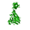
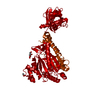
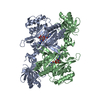
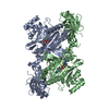
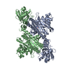
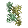
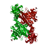
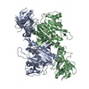
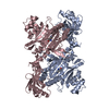
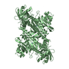
LYSYL-TRNA SYNTHETASE (LYSU) (E.C.6.1.1.6) COMPLEXED WITH LYSINE
Components
LYSYL-TRNA SYNTHETASE (LYSU)
Keywords
LIGASE (SYNTHETASE)
Function / homology
Function and homology information
RNA capping / lysine-tRNA ligase / lysine-tRNA ligase activity / lysyl-tRNA aminoacylation / tRNA aminoacylation for protein translation / ligase activity / cellular response to heat / tRNA binding / magnesium ion binding / protein homodimerization activity ...RNA capping / lysine-tRNA ligase / lysine-tRNA ligase activity / lysyl-tRNA aminoacylation / tRNA aminoacylation for protein translation / ligase activity / cellular response to heat / tRNA binding / magnesium ion binding / protein homodimerization activity / ATP binding / membrane / cytosol / cytoplasm Similarity search - Function
Bacterial/eukaryotic lysine-tRNA ligase, class II / Lysine-tRNA ligase, class II / Lysine-tRNA ligase, class II, N-terminal / Lysyl-tRNA synthetase, class II, C-terminal / Aminoacyl-tRNA synthetase, class II (D/K/N) / tRNA synthetases class II (D, K and N) / OB-fold nucleic acid binding domain, AA-tRNA synthetase-type / OB-fold nucleic acid binding domain / Bira Bifunctional Protein; Domain 2 / BirA Bifunctional Protein; domain 2 ...Bacterial/eukaryotic lysine-tRNA ligase, class II / Lysine-tRNA ligase, class II / Lysine-tRNA ligase, class II, N-terminal / Lysyl-tRNA synthetase, class II, C-terminal / Aminoacyl-tRNA synthetase, class II (D/K/N) / tRNA synthetases class II (D, K and N) / OB-fold nucleic acid binding domain, AA-tRNA synthetase-type / OB-fold nucleic acid binding domain / Bira Bifunctional Protein; Domain 2 / BirA Bifunctional Protein; domain 2 / Aminoacyl-tRNA synthetase, class II / Aminoacyl-transfer RNA synthetases class-II family profile. / Class II Aminoacyl-tRNA synthetase/Biotinyl protein ligase (BPL) and lipoyl protein ligase (LPL) / Nucleic acid-binding proteins / OB fold (Dihydrolipoamide Acetyltransferase, E2P) / Nucleic acid-binding, OB-fold / Beta Barrel / 2-Layer Sandwich / Mainly Beta / Alpha Beta Similarity search - Domain/homology
SYMMETRY THE CRYSTALLOGRAPHIC SYMMETRY TRANSFORMATIONS PRESENTED BELOW GENERATE THE SUBUNITS OF THE POLYMERIC MOLECULE. APPLIED TO RESIDUES: B 14 .. 504 CRYSTALLOGRAPHIC DYAD OPERATION SYMMETRY1 1 1.000000 0.000000 0.000000 0.00000 SYMMETRY2 1 0.000000 -1.000000 0.000000 257.80000 SYMMETRY3 1 0.000000 0.000000 -1.000000 182.08000 APPLIED TO RESIDUES: Y 1 .. 1 CRYSTALLOGRAPHIC DYAD OPERATION SYMMETRY1 2 1.000000 0.000000 0.000000 0.00000 SYMMETRY2 2 0.000000 -1.000000 0.000000 257.80000 SYMMETRY3 2 0.000000 0.000000 -1.000000 182.08000 THE ASYMMETRIC UNIT CAN ALSO BE DESCRIBED AS CONTAINING THREE SUBUNITS ARRANGED AROUND A PSEUDO 31 AXIS PARALLEL TO THE CRYSTALLOGRAPHIC C AXIS, RELATING SUBUNITS *B*, *A* AND *C'* WHERE *C'* IS CRYSTALLOGRAPHICALLY RELATED TO SUBUNIT *C*.
-
Components
#1: Protein
LYSYL-TRNASYNTHETASE (LYSU)
Mass: 57767.191 Da / Num. of mol.: 3 / Source method: isolated from a natural source / Source: (natural) Escherichia coli (E. coli) / References: UniProt: P0A8N5, lysine-tRNA ligase
In the structure databanks used in Yorodumi, some data are registered as the other names, "COVID-19 virus" and "2019-nCoV". Here are the details of the virus and the list of structure data.
Jan 31, 2019. EMDB accession codes are about to change! (news from PDBe EMDB page)
EMDB accession codes are about to change! (news from PDBe EMDB page)
The allocation of 4 digits for EMDB accession codes will soon come to an end. Whilst these codes will remain in use, new EMDB accession codes will include an additional digit and will expand incrementally as the available range of codes is exhausted. The current 4-digit format prefixed with “EMD-” (i.e. EMD-XXXX) will advance to a 5-digit format (i.e. EMD-XXXXX), and so on. It is currently estimated that the 4-digit codes will be depleted around Spring 2019, at which point the 5-digit format will come into force.
The EM Navigator/Yorodumi systems omit the EMD- prefix.
Related info.:Q: What is EMD? / ID/Accession-code notation in Yorodumi/EM Navigator
Yorodumi is a browser for structure data from EMDB, PDB, SASBDB, etc.
This page is also the successor to EM Navigator detail page, and also detail information page/front-end page for Omokage search.
The word "yorodu" (or yorozu) is an old Japanese word meaning "ten thousand". "mi" (miru) is to see.
Related info.:EMDB / PDB / SASBDB / Comparison of 3 databanks / Yorodumi Search / Aug 31, 2016. New EM Navigator & Yorodumi / Yorodumi Papers / Jmol/JSmol / Function and homology information / Changes in new EM Navigator and Yorodumi
 Movie
Movie Controller


 Open data
 Basic information
 Components
 Keywords
 Function and homology information
 X-RAY DIFFRACTION / Resolution: 2.8 Å
  Authors
 Citation
 Structure visualization
 Downloads & links
 Other downloads
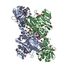

 PDBj
PDBj

 Assembly
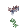

 Type: L-peptide linking / Mass: 147.195 Da / Num. of mol.: 3 / Source method: obtained synthetically / Formula: C6H15N2O2
 Mass: 18.015 Da / Num. of mol.: 375 / Source method: isolated from a natural source / Formula: H2O
 Sample preparation
 Processing