Movie
Movie Controller

[English] 日本語
 Yorodumi
- EMDB-3039: Cryo-EM structure of a mammalian ribosomal termination complex wi... -
+ Open data

- Basic information
| Entry | Database: EMDB / ID: EMD-3039 | |||||||||
|---|---|---|---|---|---|---|---|---|---|---|
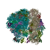
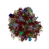
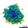
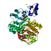
| Title | Cryo-EM structure of a mammalian ribosomal termination complex with ABCE1, eRF1(AAQ) and the UAG stop codon | |||||||||
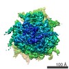
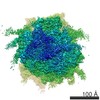
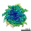
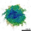
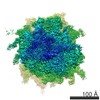
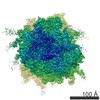
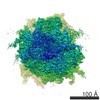
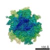
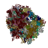
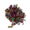
 Map data Map data | Reconstruction of a mammalian 80S ribosome-nascent chain complex containing the UAG stop codon bound to eRF1(AAQ) and ABCE1 | |||||||||
 Sample Sample |
| |||||||||
 Keywords Keywords |  ribosome / translation termination / release factor / stop codon / decoding ribosome / translation termination / release factor / stop codon / decoding | |||||||||
| Function / homology |  Function and homology information Function and homology informationnegative regulation of endoribonuclease activity / CTPase activity / translation termination factor activity / cytoplasmic translational termination / translation release factor complex / regulation of translational termination / translation release factor activity / OAS antiviral response / ribosome disassembly / translation release factor activity, codon specific ...negative regulation of endoribonuclease activity / CTPase activity / translation termination factor activity / cytoplasmic translational termination / translation release factor complex / regulation of translational termination / translation release factor activity / OAS antiviral response / ribosome disassembly / translation release factor activity, codon specific / protein methylation / ribosomal subunit / sequence-specific mRNA binding / aminoacyl-tRNA hydrolase activity / nuclear-transcribed mRNA catabolic process, nonsense-mediated decay / ribosomal subunit export from nucleus / Protein hydroxylation / ribosomal small subunit binding / Eukaryotic Translation Termination / Nonsense Mediated Decay (NMD) independent of the Exon Junction Complex (EJC) / endoribonuclease inhibitor activity / rough endoplasmic reticulum / Nonsense Mediated Decay (NMD) enhanced by the Exon Junction Complex (EJC) / rescue of stalled ribosome / translational termination / translational initiation / DNA-(apurinic or apyrimidinic site) lyase / ribonucleoside triphosphate phosphatase activity / cytosolic ribosome / Hydrolases; Acting on acid anhydrides; Acting on GTP to facilitate cellular and subcellular movement / Regulation of expression of SLITs and ROBOs / Interferon alpha/beta signaling / ribosome binding / ribosome biogenesis / regulation of translation / 4 iron, 4 sulfur cluster binding / 5S rRNA binding / ribosome / mitochondrial matrix / structural constituent of ribosome / translation / iron ion binding / ribonucleoprotein complex / GTPase activity / nucleolus / ATP hydrolysis activity / mitochondrion / RNA binding / ATP binding / membrane / cytosol / cytoplasmSimilarity search - Function | |||||||||
| Biological species |  Oryctolagus cuniculus (rabbit) / Oryctolagus cuniculus (rabbit) /  Homo sapiens (human) Homo sapiens (human) | |||||||||
| Method | single particle reconstruction / cryo EM / Resolution: 3.45 Å | |||||||||
 Authors Authors | Brown A / Shao S / Murray J / Hegde RS / Ramakrishnan V | |||||||||
 Citation Citation | Journal: Nature / Year: 2015 Title: Structural basis for stop codon recognition in eukaryotes. Authors: Alan Brown / Sichen Shao / Jason Murray / Ramanujan S Hegde / V Ramakrishnan /  Abstract: Termination of protein synthesis occurs when a translating ribosome encounters one of three universally conserved stop codons: UAA, UAG or UGA. Release factors recognize stop codons in the ribosomal ...Termination of protein synthesis occurs when a translating ribosome encounters one of three universally conserved stop codons: UAA, UAG or UGA. Release factors recognize stop codons in the ribosomal A-site to mediate release of the nascent chain and recycling of the ribosome. Bacteria decode stop codons using two separate release factors with differing specificities for the second and third bases. By contrast, eukaryotes rely on an evolutionarily unrelated omnipotent release factor (eRF1) to recognize all three stop codons. The molecular basis of eRF1 discrimination for stop codons over sense codons is not known. Here we present cryo-electron microscopy (cryo-EM) structures at 3.5-3.8 Å resolution of mammalian ribosomal complexes containing eRF1 interacting with each of the three stop codons in the A-site. Binding of eRF1 flips nucleotide A1825 of 18S ribosomal RNA so that it stacks on the second and third stop codon bases. This configuration pulls the fourth position base into the A-site, where it is stabilized by stacking against G626 of 18S rRNA. Thus, eRF1 exploits two rRNA nucleotides also used during transfer RNA selection to drive messenger RNA compaction. In this compacted mRNA conformation, stop codons are favoured by a hydrogen-bonding network formed between rRNA and essential eRF1 residues that constrains the identity of the bases. These results provide a molecular framework for eukaryotic stop codon recognition and have implications for future studies on the mechanisms of canonical and premature translation termination. | |||||||||
| History |
|
- Structure visualization
| Movie |
Movie viewer |
|---|---|
| Structure viewer | EM map: SurfViewMolmilJmol/JSmol |
| Supplemental images |
- Downloads & links
-EMDB archive
| Map data | emd_3039.map.gz | 12.7 MB | EMDB map data format | |
|---|---|---|---|---|
| Header (meta data) | emd-3039-v30.xmlemd-3039.xml | 15.2 KB 15.2 KB | Display Display | EMDB header |
| FSC (resolution estimation) | emd_3039_fsc.xml | 14.6 KB | Display | FSC data file |
| Images | emd_3039.tif | 270.8 KB | ||
| Archive directory |  http://ftp.pdbj.org/pub/emdb/structures/EMD-3039ftp://ftp.pdbj.org/pub/emdb/structures/EMD-3039 http://ftp.pdbj.org/pub/emdb/structures/EMD-3039ftp://ftp.pdbj.org/pub/emdb/structures/EMD-3039 | HTTPS FTP |
-Related structure data
| Related structure data | 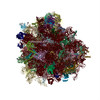 3jahMC 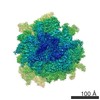 3038C 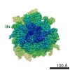 3040C  3jagC 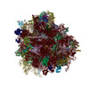 3jaiC C: citing same article ( M: atomic model generated by this map |
|---|---|
| Similar structure data |
-Links
| EMDB pages | EMDB (EBI/PDBe) / EMDataResource |
|---|---|
| Related items in Molecule of the Month |


-Map
| File | Download / File: emd_3039.map.gz / Format: CCP4 / Size: 276 MB / Type: IMAGE STORED AS FLOATING POINT NUMBER (4 BYTES) | ||||||||||||||||||||||||||||||||||||||||||||||||||||||||||||
|---|---|---|---|---|---|---|---|---|---|---|---|---|---|---|---|---|---|---|---|---|---|---|---|---|---|---|---|---|---|---|---|---|---|---|---|---|---|---|---|---|---|---|---|---|---|---|---|---|---|---|---|---|---|---|---|---|---|---|---|---|---|
| Annotation | Reconstruction of a mammalian 80S ribosome-nascent chain complex containing the UAG stop codon bound to eRF1(AAQ) and ABCE1 | ||||||||||||||||||||||||||||||||||||||||||||||||||||||||||||
| Voxel size | X=Y=Z: 1.34 Å | ||||||||||||||||||||||||||||||||||||||||||||||||||||||||||||
| Density |
| ||||||||||||||||||||||||||||||||||||||||||||||||||||||||||||
| Symmetry | Space group: 1 | ||||||||||||||||||||||||||||||||||||||||||||||||||||||||||||
| Details | EMDB XML:
CCP4 map header:
| ||||||||||||||||||||||||||||||||||||||||||||||||||||||||||||
-Supplemental data
- Sample components
-Entire : Affinity-purified 80S ribosome-nascent chain complex containing t...
| Entire | Name: Affinity-purified 80S ribosome-nascent chain complex containing the UAG stop codon bound to eRF1(AAQ) and ABCE1 |
|---|---|
| Components |
|
-Supramolecule #1000: Affinity-purified 80S ribosome-nascent chain complex containing t...
| Supramolecule | Name: Affinity-purified 80S ribosome-nascent chain complex containing the UAG stop codon bound to eRF1(AAQ) and ABCE1 type: sample / ID: 1000 / Number unique components: 6 |
|---|---|
| Molecular weight | Theoretical: 2.1 MDa |
-Supramolecule #1: 80S ribosome
| Supramolecule | Name: 80S ribosome / type: complex / ID: 1 / Recombinant expression: No / Ribosome-details: ribosome-eukaryote: ALL |
|---|---|
| Source (natural) | Organism: Oryctolagus cuniculus (rabbit) / synonym: rabbit / Cell: reticulocyte / Location in cell: cytosol |
| Molecular weight | Theoretical: 2 MDa |
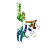
-Macromolecule #1: eukaryotic release factor 1, G183A, G184A
| Macromolecule | Name: eukaryotic release factor 1, G183A, G184A / type: protein_or_peptide / ID: 1 / Name.synonym: eRF1(AAQ) / Number of copies: 1 / Recombinant expression: Yes |
|---|---|
| Source (natural) | Organism: Homo sapiens (human) / synonym: Human |
| Recombinant expression | Organism:  Escherichia coli BL21(DE3) (bacteria) / Recombinant plasmid: pRSETA Escherichia coli BL21(DE3) (bacteria) / Recombinant plasmid: pRSETA |
| Sequence | UniProtKB: Eukaryotic peptide chain release factor subunit 1 |
-Macromolecule #2: ATP binding cassette E1
| Macromolecule | Name: ATP binding cassette E1 / type: protein_or_peptide / ID: 2 / Name.synonym: ABCE1, Rli1 / Recombinant expression: No |
|---|---|
| Source (natural) | Organism: Oryctolagus cuniculus (rabbit) / synonym: rabbit / Location in cell: cytosol |
| Sequence | UniProtKB: ATP-binding cassette sub-family E member 1 |
-Macromolecule #4: Sec61-beta
| Macromolecule | Name: Sec61-beta / type: protein_or_peptide / ID: 4 / Details: in vitro translated peptide sequence / Number of copies: 1 / Recombinant expression: No |
|---|---|
| Source (natural) | Organism: Homo sapiens (human) / synonym: Human |
-Macromolecule #3: messenger RNA
| Macromolecule | Name: messenger RNA / type: rna / ID: 3 / Name.synonym: mRNA Details: in vitro transcribed mRNA sequence containing UAA stop codon Classification: OTHER / Structure: SINGLE STRANDED / Synthetic?: No |
|---|---|
| Source (natural) | Organism: Homo sapiens (human) / synonym: Human |
| Sequence | String: UCAAAGUUUA GG |
-Macromolecule #5: transfer RNA
| Macromolecule | Name: transfer RNA / type: rna / ID: 5 / Name.synonym: tRNA / Classification: OTHER / Structure: OTHER / Synthetic?: No |
|---|---|
| Source (natural) | Organism: Oryctolagus cuniculus (rabbit) / synonym: rabbit |
| Molecular weight | Theoretical: 20 KDa |
-Experimental details
-Structure determination
| Method | cryo EM |
|---|---|
 Processing Processing | single particle reconstruction |
| Aggregation state | particle |
-Sample preparation
| Buffer | pH: 7.4 / Details: 50 mM Hepes, 100 mM KAc, 5 mM MgAc2, 1 mM DTT |
|---|---|
| Grid | Details: R2/2 400 mesh Cu grids with thin continuous carbon support, glow discharged |
| Vitrification | Cryogen name: ETHANE / Chamber humidity: 100 % / Instrument: FEI VITROBOT MARK III / Method: 30 sec wait time, blot for 3 sec before plunging |
- Electron microscopy
| Microscope | FEI TITAN KRIOS |
|---|---|
| Electron beam | Acceleration voltage: 300 kV / Electron source: FIELD EMISSION GUN |
| Electron optics | Calibrated magnification: 104478 / Illumination mode: FLOOD BEAM / Imaging mode: BRIGHT FIELDBright-field microscopy / Cs: 2.7 mm / Nominal defocus max: 3.6 µm / Nominal defocus min: 1.7 µm / Nominal magnification: 59000 |
| Specialist optics | Energy filter - Name: FEI |
| Sample stage | Specimen holder model: FEI TITAN KRIOS AUTOGRID HOLDER |
| Details | Automated data acquisition using EPU (FEI) |
| Date | Feb 25, 2015 |
| Image recording | Category: CCD / Film or detector model: FEI FALCON II (4k x 4k) / Number real images: 1601 / Average electron dose: 30 e/Å2 Details: Every image is the average of 17 frames recorded by the direct electron detector |
| Experimental equipment |  Model: Titan Krios / Image courtesy: FEI Company |
-Image processing
| Final reconstruction | Applied symmetry - Point group: C1 (asymmetric) / Resolution.type: BY AUTHOR / Resolution: 3.45 Å / Resolution method: OTHER / Software - Name: CTFFIND3, RELION / Number images used: 20515 |
|---|---|
| FSC plot (resolution estimation) | 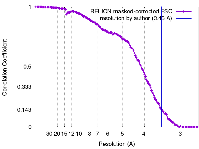 |
-Atomic model buiding 1
| Initial model | PDB ID: |
|---|---|
| Software | Name: Chimera, Coot |
| Refinement | Space: RECIPROCAL / Protocol: FLEXIBLE FIT |
| Output model | PDB-3jah: |
-Atomic model buiding 2
| Initial model | PDB ID: |
|---|---|
| Software | Name: Chimera, Coot |
| Refinement | Space: RECIPROCAL / Protocol: FLEXIBLE FIT |
| Output model | PDB-3jah: |

-Atomic model buiding 3
| Initial model | PDB ID: |
|---|---|
| Software | Name: Chimera, Coot |
| Refinement | Space: RECIPROCAL / Protocol: FLEXIBLE FIT |
| Output model | PDB-3jah: |
-Atomic model buiding 4
| Initial model | PDB ID: |
|---|---|
| Software | Name: Chimera, Coot |
| Details | Sequence was modified in Coot to agree with rabbit sequence |
| Refinement | Space: RECIPROCAL / Protocol: FLEXIBLE FIT |
| Output model | PDB-3jah: |
-Atomic model buiding 5
| Initial model | PDB ID: |
|---|---|
| Software | Name: Chimera, Coot |
| Details | Sequence was modified in Coot to agree with the most prevalent tRNA sequence for each particular codon |
| Refinement | Space: RECIPROCAL / Protocol: FLEXIBLE FIT |
| Output model | PDB-3jah: |