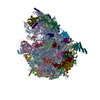
 Movie
Movie Controller

[English] 日本語
 Yorodumi
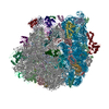
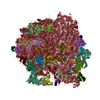
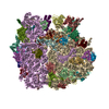
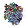
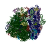
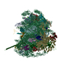
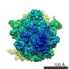
- PDB-3j92: Structure and assembly pathway of the ribosome quality control complex -
+ Open data

- Basic information
| Entry | Database: PDB / ID: 3j92 | ||||||
|---|---|---|---|---|---|---|---|
| Title | Structure and assembly pathway of the ribosome quality control complex | ||||||
 Components Components |
| ||||||
 Keywords Keywords | RIBOSOME/LIGASE / RWD / RING / quality control / RIBOSOME-LIGASE complex | ||||||
| Function / homology |  Function and homology information Function and homology informationalpha-aminoacyl-tRNA binding / CAT tailing / RQC complex / nuclear export / ribosome-associated ubiquitin-dependent protein catabolic process / ribosomal large subunit binding / protein autoubiquitination / rescue of stalled cytosolic ribosome / cytosolic ribosome / RING-type E3 ubiquitin transferase ...alpha-aminoacyl-tRNA binding / CAT tailing / RQC complex / nuclear export / ribosome-associated ubiquitin-dependent protein catabolic process / ribosomal large subunit binding / protein autoubiquitination / rescue of stalled cytosolic ribosome / cytosolic ribosome / RING-type E3 ubiquitin transferase / ubiquitin protein ligase activity / Antigen processing: Ubiquitination & Proteasome degradation / protein-containing complex assembly / tRNA binding / zinc ion binding / nucleus / cytosol Similarity search - Function | ||||||
| Biological species |  Homo sapiens (human) Homo sapiens (human) | ||||||
| Method | ELECTRON MICROSCOPY / single particle reconstruction / cryo EM / Resolution: 3.6 Å | ||||||
 Authors Authors | Shao, S. / Brown, A. / Santhanam, B. / Hegde, R.S. | ||||||
 Citation Citation | Journal: Mol Cell / Year: 2015 Title: Structure and assembly pathway of the ribosome quality control complex. Authors: Sichen Shao / Alan Brown / Balaji Santhanam / Ramanujan S Hegde /  Abstract: During ribosome-associated quality control, stalled ribosomes are split into subunits and the 60S-housed nascent polypeptides are poly-ubiquitinated by Listerin. How this low-abundance ubiquitin ...During ribosome-associated quality control, stalled ribosomes are split into subunits and the 60S-housed nascent polypeptides are poly-ubiquitinated by Listerin. How this low-abundance ubiquitin ligase targets rare stall-generated 60S among numerous empty 60S is unknown. Here, we show that Listerin specificity for nascent chain-60S complexes depends on nuclear export mediator factor (NEMF). The 3.6 Å cryo-EM structure of a nascent chain-containing 60S-Listerin-NEMF complex revealed that NEMF makes multiple simultaneous contacts with 60S and peptidyl-tRNA to sense nascent chain occupancy. Structural and mutational analyses showed that ribosome-bound NEMF recruits and stabilizes Listerin's N-terminal domain, while Listerin's C-terminal RWD domain directly contacts the ribosome to position the adjacent ligase domain near the nascent polypeptide exit tunnel. Thus, highly specific nascent chain targeting by Listerin is imparted by the avidity gained from a multivalent network of context-specific individually weak interactions, highlighting a new principle of client recognition during protein quality control. | ||||||
| History |
|
- Structure visualization
| Movie |
Movie viewer |
|---|---|
| Structure viewer | Molecule: MolmilJmol/JSmol |
 UCSF Chimera
- Downloads & links
-Download
| PDBx/mmCIF format | 3j92.cif.gz | 3.6 MB | Display | PDBx/mmCIF format |
|---|---|---|---|---|
| PDB format | pdb3j92.ent.gz | Display | PDB format | |
| PDBx/mmJSON format | 3j92.json.gz | Tree view | PDBx/mmJSON format | |
| Others |  Other downloads Other downloads |
-Validation report
| Arichive directory | https://data.pdbj.org/pub/pdb/validation_reports/j9/3j92ftp://data.pdbj.org/pub/pdb/validation_reports/j9/3j92 | HTTPS FTP |
|---|
-Related structure data
| Related structure data |  2832MC M: map data used to model this data C: citing same article ( |
|---|---|
| Similar structure data |

-Links
 PDBj
PDBj


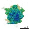
- Assembly
| Deposited unit | 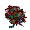
|
|---|---|
| 1 |
|
-Components
+Protein , 47 types, 51 molecules ABCDEFGHIJLMNOPQRSTUVWXYZabcde...
-RNA chain , 4 types, 4 molecules 2578
| #48: RNA chain | Mass: 24816.811 Da / Num. of mol.: 1 / Source method: isolated from a natural source / Source: (natural) |
|---|---|
| #49: RNA chain | Mass: 1187230.000 Da / Num. of mol.: 1 / Source method: isolated from a natural source / Source: (natural) |
| #50: RNA chain | Mass: 38691.914 Da / Num. of mol.: 1 / Source method: isolated from a natural source / Source: (natural) |
| #51: RNA chain | Mass: 50143.648 Da / Num. of mol.: 1 / Source method: isolated from a natural source / Source: (natural) |
-Non-polymers , 2 types, 164 molecules 

| #52: Chemical | ChemComp-MG /  Mass: 24.305 Da / Num. of mol.: 159 / Source method: obtained synthetically / Formula: Mg Mass: 24.305 Da / Num. of mol.: 159 / Source method: obtained synthetically / Formula: Mg#53: Chemical | ChemComp-ZN /  Mass: 65.409 Da / Num. of mol.: 5 / Source method: obtained synthetically / Formula: Zn Mass: 65.409 Da / Num. of mol.: 5 / Source method: obtained synthetically / Formula: Zn |
|---|
-Details
| Has protein modification | Y |
|---|---|
| Sequence details | ALTHOUGH ONLY ONE INSTANCE EACH OF PROTEINS NEMF AND LISTERIN IS PRESENT WITHIN THE COMPLEX, EACH ...ALTHOUGH ONLY ONE INSTANCE EACH OF PROTEINS NEMF AND LISTERIN IS PRESENT WITHIN THE COMPLEX, EACH OF THESE PROTEINS IS REPRESENTE |
-Experimental details
-Experiment
| Experiment | Method: ELECTRON MICROSCOPY |
|---|---|
| EM experiment | Aggregation state: PARTICLE / 3D reconstruction method: single particle reconstruction |
- Sample preparation
| Component |
| ||||||||||||||||||||||||||||
|---|---|---|---|---|---|---|---|---|---|---|---|---|---|---|---|---|---|---|---|---|---|---|---|---|---|---|---|---|---|
| Buffer solution | Name: 50 mM HEPES, 100 mM potassium acetate, 5 mM magnesium acetate, 1 mM DTT pH: 7.4 Details: 50 mM HEPES, 100 mM potassium acetate, 5 mM magnesium acetate, 1 mM DTT | ||||||||||||||||||||||||||||
| Specimen | Conc.: 0.3 mg/ml / Embedding applied: NO / Shadowing applied: NO / Staining applied: NO / Vitrification applied: YES | ||||||||||||||||||||||||||||
| Specimen support | Details: Quantifoil R2/2 on 400 mesh Cu grid with thin carbon support | ||||||||||||||||||||||||||||
| Vitrification | Instrument: FEI VITROBOT MARK I / Cryogen name: ETHANE / Humidity: 100 % Details: Blot for 3 seconds before plunging into liquid ethane (FEI VITROBOT MARK I). Method: Blot for 3 seconds before plunging |
- Electron microscopy imaging
| Experimental equipment |  Model: Titan Krios / Image courtesy: FEI Company | ||||||
|---|---|---|---|---|---|---|---|
| EM imaging | Accelerating voltage: 300 kV / Calibrated magnification: 104478 X / Details: Objective lens astigmatism was corrected at 59,000 times magnification. / Electron source:
| ||||||
| Image recording | Electron dose: 30 e/Å2 / Film or detector model: FEI FALCON II (4k x 4k) |
- Processing
| Software | Name: REFMAC / Version: 5.8.0091 2014/10/05 / Classification: refinement / Contact author: Garib N. Murshudov / Contact author email: garib[at]mrc-lmb.cam.ac.uk Description: (un)restrained refinement or idealisation of macromolecular structures | |||||||||||||||||||||
|---|---|---|---|---|---|---|---|---|---|---|---|---|---|---|---|---|---|---|---|---|---|---|
| EM software |
| |||||||||||||||||||||
| Symmetry | Point symmetry: C1 (asymmetric) | |||||||||||||||||||||
| 3D reconstruction | Resolution: 3.6 Å / Resolution method: FSC 0.143 CUT-OFF / Num. of particles: 63826 / Nominal pixel size: 1.34 Å / Actual pixel size: 1.34 Å / Symmetry type: POINT | |||||||||||||||||||||
| Atomic model building | Protocol: RIGID BODY FIT / Space: RECIPROCAL / Details: REFINEMENT PROTOCOL--rigid body | |||||||||||||||||||||
| Atomic model building |
| |||||||||||||||||||||
| Refinement | Details: Hydrogens have been added in their riding positions | |||||||||||||||||||||
| Refinement step | Cycle: LAST
|

