Movie
Movie Controller

[English] 日本語
 Yorodumi
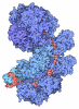
- PDB-3jag: Structure of a mammalian ribosomal termination complex with ABCE1... -
+ Open data

- Basic information
| Entry | Database: PDB / ID: 3jag | ||||||
|---|---|---|---|---|---|---|---|
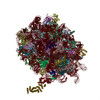
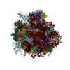
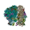
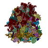
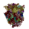
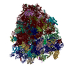
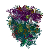
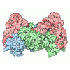
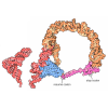
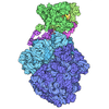
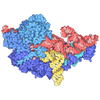
| Title | Structure of a mammalian ribosomal termination complex with ABCE1, eRF1(AAQ), and the UAA stop codon | ||||||
 Components Components |
| ||||||
 Keywords Keywords | RIBOSOME / termination / eRF1 / ABCE1 | ||||||
| Function / homology |  Function and homology information Function and homology informationtranslation termination factor activity / translation release factor complex / cytoplasmic translational termination / regulation of translational termination / translation release factor activity / translation release factor activity, codon specific / sequence-specific mRNA binding / peptidyl-tRNA hydrolase activity / protein methylation / regulation of translation involved in cellular response to UV ...translation termination factor activity / translation release factor complex / cytoplasmic translational termination / regulation of translational termination / translation release factor activity / translation release factor activity, codon specific / sequence-specific mRNA binding / peptidyl-tRNA hydrolase activity / protein methylation / regulation of translation involved in cellular response to UV / positive regulation of intrinsic apoptotic signaling pathway in response to DNA damage by p53 class mediator / protein-DNA complex disassembly / positive regulation of DNA damage response, signal transduction by p53 class mediator / Protein hydroxylation / Eukaryotic Translation Termination / ubiquitin ligase inhibitor activity / 90S preribosome / Nonsense Mediated Decay (NMD) independent of the Exon Junction Complex (EJC) / positive regulation of signal transduction by p53 class mediator / translational termination / phagocytic cup / Nonsense Mediated Decay (NMD) enhanced by the Exon Junction Complex (EJC) / negative regulation of ubiquitin-dependent protein catabolic process / translation regulator activity / ribosomal small subunit export from nucleus / rough endoplasmic reticulum / positive regulation of apoptotic signaling pathway / MDM2/MDM4 family protein binding / DNA-(apurinic or apyrimidinic site) lyase / class I DNA-(apurinic or apyrimidinic site) endonuclease activity / ribosomal large subunit biogenesis / maturation of SSU-rRNA from tricistronic rRNA transcript (SSU-rRNA, 5.8S rRNA, LSU-rRNA) / DNA damage response, signal transduction by p53 class mediator / maturation of LSU-rRNA from tricistronic rRNA transcript (SSU-rRNA, 5.8S rRNA, LSU-rRNA) / maturation of SSU-rRNA / positive regulation of translation / small-subunit processome / cellular response to gamma radiation / spindle / Regulation of expression of SLITs and ROBOs / mRNA 5'-UTR binding / transcription coactivator binding / rRNA processing / cytosolic ribosome / glucose homeostasis / ribosomal small subunit assembly / ribosome biogenesis / ribosome binding / ribosomal small subunit biogenesis / small ribosomal subunit rRNA binding / 5S rRNA binding / ribosomal large subunit assembly / small ribosomal subunit / cytosolic small ribosomal subunit / large ribosomal subunit rRNA binding / cytosolic large ribosomal subunit / perikaryon / cytoplasmic translation / protein stabilization / mitochondrial inner membrane / rRNA binding / postsynaptic density / ribonucleoprotein complex / ribosome / translation / structural constituent of ribosome / mRNA binding / ubiquitin protein ligase binding / centrosome / positive regulation of cell population proliferation / nucleolus / dendrite / synapse / perinuclear region of cytoplasm / negative regulation of transcription by RNA polymerase II / endoplasmic reticulum / ATP hydrolysis activity / RNA binding / zinc ion binding / nucleoplasm / ATP binding / membrane / nucleus / plasma membrane / cytosol / cytoplasm Similarity search - Function | ||||||
| Biological species |  Homo sapiens (human) Homo sapiens (human)  Oryctolagus cuniculus (rabbit) Oryctolagus cuniculus (rabbit) | ||||||
| Method | ELECTRON MICROSCOPY / single particle reconstruction / cryo EM / Resolution: 3.65 Å | ||||||
 Authors Authors | Brown, A. / Shao, S. / Murray, J. / Hegde, R.S. / Ramakrishnan, V. | ||||||
 Citation Citation | Journal: Nature / Year: 2015 Title: Structural basis for stop codon recognition in eukaryotes. Authors: Alan Brown / Sichen Shao / Jason Murray / Ramanujan S Hegde / V Ramakrishnan /  Abstract: Termination of protein synthesis occurs when a translating ribosome encounters one of three universally conserved stop codons: UAA, UAG or UGA. Release factors recognize stop codons in the ribosomal ...Termination of protein synthesis occurs when a translating ribosome encounters one of three universally conserved stop codons: UAA, UAG or UGA. Release factors recognize stop codons in the ribosomal A-site to mediate release of the nascent chain and recycling of the ribosome. Bacteria decode stop codons using two separate release factors with differing specificities for the second and third bases. By contrast, eukaryotes rely on an evolutionarily unrelated omnipotent release factor (eRF1) to recognize all three stop codons. The molecular basis of eRF1 discrimination for stop codons over sense codons is not known. Here we present cryo-electron microscopy (cryo-EM) structures at 3.5-3.8 Å resolution of mammalian ribosomal complexes containing eRF1 interacting with each of the three stop codons in the A-site. Binding of eRF1 flips nucleotide A1825 of 18S ribosomal RNA so that it stacks on the second and third stop codon bases. This configuration pulls the fourth position base into the A-site, where it is stabilized by stacking against G626 of 18S rRNA. Thus, eRF1 exploits two rRNA nucleotides also used during transfer RNA selection to drive messenger RNA compaction. In this compacted mRNA conformation, stop codons are favoured by a hydrogen-bonding network formed between rRNA and essential eRF1 residues that constrains the identity of the bases. These results provide a molecular framework for eukaryotic stop codon recognition and have implications for future studies on the mechanisms of canonical and premature translation termination. | ||||||
| History |
|
- Structure visualization
| Movie |
Movie viewer |
|---|---|
| Structure viewer | Molecule: MolmilJmol/JSmol |
 UCSF Chimera
- Downloads & links
-Download
| PDBx/mmCIF format | 3jag.cif.gz | 5.6 MB | Display | PDBx/mmCIF format |
|---|---|---|---|---|
| PDB format | pdb3jag.ent.gz | Display | PDB format | |
| PDBx/mmJSON format | 3jag.json.gz | Tree view | PDBx/mmJSON format | |
| Others |  Other downloads Other downloads |
-Validation report
| Arichive directory | https://data.pdbj.org/pub/pdb/validation_reports/ja/3jagftp://data.pdbj.org/pub/pdb/validation_reports/ja/3jag | HTTPS FTP |
|---|
-Related structure data
| Related structure data | 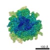 3038MC 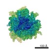 3039C 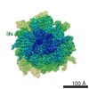 3040C  3jahC  3jaiC M: map data used to model this data C: citing same article ( |
|---|---|
| Similar structure data |
-Links
 PDBj
PDBj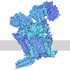
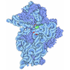

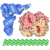
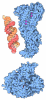
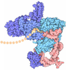
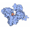
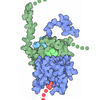


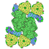


- Assembly
| Deposited unit | 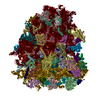
|
|---|---|
| 1 |
|
-Components
+Protein , 77 types, 77 molecules ABCDEFGHIJLMNOPQRSTUVWXYZabcde...

 Escherichia coli BL21(DE3) (bacteria)
-Protein/peptide , 3 types, 3 molecules ln1
| #37: Protein/peptide | Mass: 6295.562 Da / Num. of mol.: 1 / Source method: isolated from a natural source / Source: (natural) Oryctolagus cuniculus (rabbit) / Cell: reticulocyte lysate / References: UniProt: G1SYU7 |
|---|---|
| #39: Protein/peptide | Mass: 3213.075 Da / Num. of mol.: 1 / Source method: isolated from a natural source / Source: (natural) Oryctolagus cuniculus (rabbit) / Cell: reticulocyte lysate / References: UniProt: A0A087WNH4 |
| #45: Protein/peptide | Mass: 1788.032 Da / Num. of mol.: 1 / Source method: isolated from a natural source / Source: (natural) Oryctolagus cuniculus (rabbit) / Cell: reticulocyte lysate |
-RNA chain , 7 types, 7 molecules 235789hh
| #46: RNA chain | Mass: 24436.551 Da / Num. of mol.: 1 / Source method: isolated from a natural source / Source: (natural) Oryctolagus cuniculus (rabbit) / Cell: reticulocyte lysate |
|---|---|
| #47: RNA chain | Mass: 24102.275 Da / Num. of mol.: 1 / Source method: isolated from a natural source / Source: (natural) Oryctolagus cuniculus (rabbit) / Cell: reticulocyte lysate |
| #48: RNA chain | Mass: 1186579.500 Da / Num. of mol.: 1 / Source method: isolated from a natural source / Source: (natural) Oryctolagus cuniculus (rabbit) / Cell: reticulocyte lysate |
| #49: RNA chain | Mass: 38691.914 Da / Num. of mol.: 1 / Source method: isolated from a natural source / Source: (natural) Oryctolagus cuniculus (rabbit) / Cell: reticulocyte lysate |
| #50: RNA chain | Mass: 50143.648 Da / Num. of mol.: 1 / Source method: isolated from a natural source / Source: (natural) Oryctolagus cuniculus (rabbit) / Cell: reticulocyte lysate |
| #51: RNA chain | Mass: 554751.312 Da / Num. of mol.: 1 / Source method: isolated from a natural source / Source: (natural) Oryctolagus cuniculus (rabbit) / Cell: reticulocyte lysate |
| #85: RNA chain | Mass: 3821.328 Da / Num. of mol.: 1 / Source method: isolated from a natural source / Source: (natural) Oryctolagus cuniculus (rabbit) / Cell: reticulocyte lysate |
-Non-polymers , 4 types, 207 molecules 


| #88: Chemical | ChemComp-MG /  Mass: 24.305 Da / Num. of mol.: 195 / Source method: obtained synthetically / Formula: Mg Mass: 24.305 Da / Num. of mol.: 195 / Source method: obtained synthetically / Formula: Mg#89: Chemical | ChemComp-ZN /  Mass: 65.409 Da / Num. of mol.: 8 / Source method: obtained synthetically / Formula: Zn Mass: 65.409 Da / Num. of mol.: 8 / Source method: obtained synthetically / Formula: Zn#90: Chemical | 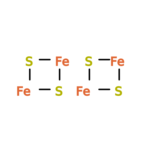 Mass: 351.640 Da / Num. of mol.: 2 / Source method: obtained synthetically / Formula: Fe4S4 Mass: 351.640 Da / Num. of mol.: 2 / Source method: obtained synthetically / Formula: Fe4S4#91: Chemical | 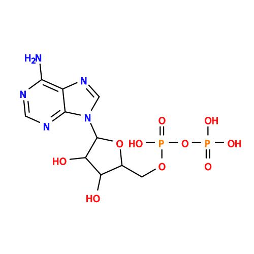 Mass: 427.201 Da / Num. of mol.: 2 / Source method: obtained synthetically / Formula: C10H15N5O10P2 / Comment: ADP, energy-carrying molecule*YM Mass: 427.201 Da / Num. of mol.: 2 / Source method: obtained synthetically / Formula: C10H15N5O10P2 / Comment: ADP, energy-carrying molecule*YM |
|---|
-Details
| Has protein modification | Y |
|---|
-Experimental details
-Experiment
| Experiment | Method: ELECTRON MICROSCOPY |
|---|---|
| EM experiment | Aggregation state: PARTICLE / 3D reconstruction method: single particle reconstruction |
- Sample preparation
| Component |
| ||||||||||||||||||||||||||||||||||||||||
|---|---|---|---|---|---|---|---|---|---|---|---|---|---|---|---|---|---|---|---|---|---|---|---|---|---|---|---|---|---|---|---|---|---|---|---|---|---|---|---|---|---|
| Buffer solution | Name: 50 mM HEPES, 100 mM potassium acetate, 5 mM magnesium acetate, 1 mM DTT pH: 7.4 Details: 50 mM HEPES, 100 mM potassium acetate, 5 mM magnesium acetate, 1 mM DTT | ||||||||||||||||||||||||||||||||||||||||
| Specimen | Embedding applied: NO / Shadowing applied: NO / Staining applied: NO / Vitrification applied: YES | ||||||||||||||||||||||||||||||||||||||||
| Specimen support | Details: Quantifoil R2/2 400 mesh Cu grid with thin continuous carbon support, glow discharged | ||||||||||||||||||||||||||||||||||||||||
| Vitrification | Instrument: FEI VITROBOT MARK III / Cryogen name: ETHANE / Humidity: 100 % Details: After 30 second wait time, blot for 3 seconds before plunging into liquid ethane (FEI VITROBOT MARK III). Method: After 30 second wait time, blot for 3 seconds before plunging |
- Electron microscopy imaging
| Experimental equipment |  Model: Titan Krios / Image courtesy: FEI Company | ||||||
|---|---|---|---|---|---|---|---|
| EM imaging | Accelerating voltage: 300 kV / Calibrated magnification: 104478 X / Details: Automated data acquisition using EPU (FEI) / Electron source:
| ||||||
| Image recording | Electron dose: 30 e/Å2 / Film or detector model: FEI FALCON II (4k x 4k) | ||||||
| Image scans | Num. digital images: 2672 |
- Processing
| EM software |
| ||||||||||||||||||||||||||||||||||||||||||
|---|---|---|---|---|---|---|---|---|---|---|---|---|---|---|---|---|---|---|---|---|---|---|---|---|---|---|---|---|---|---|---|---|---|---|---|---|---|---|---|---|---|---|---|
| Symmetry | Point symmetry: C1 (asymmetric) | ||||||||||||||||||||||||||||||||||||||||||
| 3D reconstruction | Resolution: 3.65 Å / Resolution method: FSC 0.143 CUT-OFF / Num. of particles: 49979 / Nominal pixel size: 1.34 Å / Actual pixel size: 1.34 Å / Symmetry type: POINT | ||||||||||||||||||||||||||||||||||||||||||
| Atomic model building |
| ||||||||||||||||||||||||||||||||||||||||||
| Atomic model building |
| ||||||||||||||||||||||||||||||||||||||||||
| Refinement step | Cycle: LAST
|