 Movie
Movie Controller
[English] 日本語
 Yorodumi
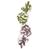
- SASDBD4: Plakin domain fragment of Human desmoplakin (C-terminal region sp... -
+ Open data

- Basic information
| Entry | Database: SASBDB / ID: SASDBD4 |
|---|---|
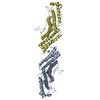
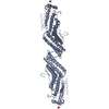
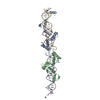
 Sample Sample | Plakin domain fragment of Human desmoplakin (C-terminal region spectrin repeats: SR7-SR8-SR9)
|
| Function / homology |  Function and homology information Function and homology informationbundle of His cell-Purkinje myocyte adhesion involved in cell communication / cell adhesive protein binding involved in bundle of His cell-Purkinje myocyte communication / desmosome organization / Keratinization / peptide cross-linking / protein localization to cell-cell junction / epithelial cell-cell adhesion / intermediate filament organization / ventricular compact myocardium morphogenesis / desmosome ...bundle of His cell-Purkinje myocyte adhesion involved in cell communication / cell adhesive protein binding involved in bundle of His cell-Purkinje myocyte communication / desmosome organization / Keratinization / peptide cross-linking / protein localization to cell-cell junction / epithelial cell-cell adhesion / intermediate filament organization / ventricular compact myocardium morphogenesis / desmosome / fascia adherens / intermediate filament cytoskeleton organization / Formation of the cornified envelope / cornified envelope / adherens junction organization / Apoptotic cleavage of cell adhesion proteins / regulation of ventricular cardiac muscle cell action potential / RND1 GTPase cycle / RND3 GTPase cycle / intermediate filament / skin development / regulation of heart rate by cardiac conduction / ficolin-1-rich granule membrane / intercalated disc / epidermis development / keratinocyte differentiation / protein kinase C binding / adherens junction / wound healing / cell-cell adhesion / structural constituent of cytoskeleton / scaffold protein binding / basolateral plasma membrane / Neutrophil degranulation / structural molecule activity / RNA binding / extracellular exosome / nucleus / plasma membrane / cytoplasm Similarity search - Function |
| Biological species |  Homo sapiens (human) Homo sapiens (human) |
 Citation Citation | Journal: J Biol Chem / Year: 2016 Title: The Structure of the Plakin Domain of Plectin Reveals an Extended Rod-like Shape. Authors: Esther Ortega / José A Manso / Rubén M Buey / Ana M Carballido / Arturo Carabias / Arnoud Sonnenberg / José M de Pereda /   Abstract: Plakins are large multi-domain proteins that interconnect cytoskeletal structures. Plectin is a prototypical plakin that tethers intermediate filaments to membrane-associated complexes. Most plakins ...Plakins are large multi-domain proteins that interconnect cytoskeletal structures. Plectin is a prototypical plakin that tethers intermediate filaments to membrane-associated complexes. Most plakins contain a plakin domain formed by up to nine spectrin repeats (SR1-SR9) and an SH3 domain. The plakin domains of plectin and other plakins harbor binding sites for junctional proteins. We have combined x-ray crystallography with small angle x-ray scattering (SAXS) to elucidate the structure of the plakin domain of plectin, extending our previous analysis of the SR1 to SR5 region. Two crystal structures of the SR5-SR6 region allowed us to characterize its uniquely wide inter-repeat conformational variability. We also report the crystal structures of the SR7-SR8 region, refined to 1.8 Å, and the SR7-SR9 at lower resolution. The SR7-SR9 region, which is conserved in all other plakin domains, forms a rigid segment stabilized by uniquely extensive inter-repeat contacts mediated by unusually long helices in SR8 and SR9. Using SAXS we show that in solution the SR3-SR6 and SR7-SR9 regions are rod-like segments and that SR3-SR9 of plectin has an extended shape with a small central kink. Other plakins, such as bullous pemphigoid antigen 1 and microtubule and actin cross-linking factor 1, are likely to have similar extended plakin domains. In contrast, desmoplakin has a two-segment structure with a central flexible hinge. The continuous versus segmented structures of the plakin domains of plectin and desmoplakin give insight into how different plakins might respond to tension and transmit mechanical signals. |
 Contact author Contact author |
|
- Structure visualization
| Structure viewer | Molecule: MolmilJmol/JSmol |
|---|
- Downloads & links
-Data source
| SASBDB page |  SASDBD4 SASDBD4 |
|---|
-Related structure data


-External links
| Related items in Molecule of the Month |
|---|


-Models
| Model #497 |   Type: dummy / Software: DAMMIF / Radius of dummy atoms: 3.50 A / Chi-square value: 1.393 / P-value: 0.415500  Search similar-shape structures of this assembly by Omokage search (details) Search similar-shape structures of this assembly by Omokage search (details) |
|---|
-Sample
| Sample | Name: Plakin domain fragment of Human desmoplakin (C-terminal region spectrin repeats: SR7-SR8-SR9) Specimen concentration: 0.96-7.70 |
|---|---|
| Buffer | Name: sodium phosphate / Concentration: 20.00 mM / pH: 7.5 / Composition: 150 mM NaCl, 5% glycerol, 3 mM DTT |
| Entity #322 | Name: Desmoplakin / Type: protein Description: Plakin domain fragment of Human Desmoplakin encompassing spectrin repeats SR7-SR8-SR9 Formula weight: 41.982 / Num. of mol.: 1 / Source: Homo sapiens / References: UniProt: P15924 Sequence: GSHMENDKQE TWMLMELQKI RRQIEHCEGR MTLKNLPLAD QGSSHHITVK INELKSVQND SQAIAEVLNQ LKDMLANFRG SEKYCYLQNE VFGLFQKLEN INGVTDGYLN SLCTVRALLQ AILQTEDMLK VYEARLTEEE TVCLDLDKVE AYRCGLKKIK NDLNLKKSLL ...Sequence: GSHMENDKQE TWMLMELQKI RRQIEHCEGR MTLKNLPLAD QGSSHHITVK INELKSVQND SQAIAEVLNQ LKDMLANFRG SEKYCYLQNE VFGLFQKLEN INGVTDGYLN SLCTVRALLQ AILQTEDMLK VYEARLTEEE TVCLDLDKVE AYRCGLKKIK NDLNLKKSLL ATMKTELQKA QQIHSQTSQQ YPLYDLDLGK FGEKVTQLTD RWQRIDKQID FRLWDLEKQI KQLRNYRDNY QAFCKWLYDA KRRQDSLESM KFGDSNTVMR FLNEQKNLHS EISGKRDKSE EVQKIAELCA NSIKDYELQL ASYTSGLETL LNIPIKRTMI QSPSGVILQE AADVHARYIE LLTRSGDYYR |
-Experimental information
| Beam | Instrument name: PETRA III P12 / City: Hamburg / 国: Germany  / Type of source: X-ray synchrotron / Wavelength: 0.12 Å / Dist. spec. to detc.: 3.1 mm / Type of source: X-ray synchrotron / Wavelength: 0.12 Å / Dist. spec. to detc.: 3.1 mm | ||||||||||||||||||
|---|---|---|---|---|---|---|---|---|---|---|---|---|---|---|---|---|---|---|---|
| Detector | Name: Pilatus 2M | ||||||||||||||||||
| Scan | 
| ||||||||||||||||||
| Distance distribution function P(R) | 
| ||||||||||||||||||
| Result | 
|
