 Movie
Movie Controller

[English] 日本語
 Yorodumi
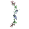
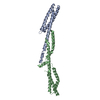
- PDB-6iei: Loop deletion and proline insertion mutant (deleting six residues... -
+ Open data

- Basic information
| Entry | Database: PDB / ID: 6iei | ||||||
|---|---|---|---|---|---|---|---|
| Title | Loop deletion and proline insertion mutant (deleting six residues and inserted five proline residues) | ||||||
 Components Components | Outer surface protein A | ||||||
 Keywords Keywords | DE NOVO PROTEIN / Outer surface protein A / OspA / LIPID BINDING PROTEIN | ||||||
| Function / homology | Outer surface lipoprotein, Borrelia / Outer surface lipoprotein domain superfamily / Borrelia lipoprotein / cell outer membrane / Prokaryotic membrane lipoprotein lipid attachment site profile. / cell surface / membrane / Outer surface protein A Function and homology information Function and homology information | ||||||
| Biological species |  Borrelia burgdorferi B31 (bacteria) Borrelia burgdorferi B31 (bacteria) | ||||||
| Method |  X-RAY DIFFRACTION / SYNCHROTRON / MOLECULAR REPLACEMENT / Resolution: 2.4 Å X-RAY DIFFRACTION / SYNCHROTRON / MOLECULAR REPLACEMENT / Resolution: 2.4 Å | ||||||
 Authors Authors | Shiga, S. / Makabe, K. | ||||||
 Citation Citation | Journal: Chembiochem / Year: 2019 Title: Domain-Swapping Design by Polyproline Rod Insertion. Authors: Shiga, S. / Yamanaka, M. / Fujiwara, W. / Hirota, S. / Goda, S. / Makabe, K. | ||||||
| History |
|
- Structure visualization
| Structure viewer | Molecule: MolmilJmol/JSmol |
|---|
- Downloads & links
-Download
| PDBx/mmCIF format | 6iei.cif.gz | 61.3 KB | Display | PDBx/mmCIF format |
|---|---|---|---|---|
| PDB format | pdb6iei.ent.gz | 42.9 KB | Display | PDB format |
| PDBx/mmJSON format | 6iei.json.gz | Tree view | PDBx/mmJSON format | |
| Others |  Other downloads Other downloads |
-Validation report
| Arichive directory | https://data.pdbj.org/pub/pdb/validation_reports/ie/6ieiftp://data.pdbj.org/pub/pdb/validation_reports/ie/6iei | HTTPS FTP |
|---|
-Related structure data
| Related structure data |  6aisC  6icsC  6idcC  6iysC  2g8cS S: Starting model for refinement C: citing same article ( |
|---|---|
| Similar structure data |


-Links
 PDBj
PDBj


- Assembly
| Deposited unit | 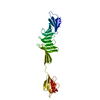
| ||||||||
|---|---|---|---|---|---|---|---|---|---|
| 1 | 
| ||||||||
| Unit cell |
|
-Components
| #1: Protein | Mass: 26348.541 Da / Num. of mol.: 1 Source method: isolated from a genetically manipulated source Source: (gene. exp.) Borrelia burgdorferi B31 (bacteria) / Strain: ATCC 35210 / B31 / CIP 102532 / DSM 4680 / Gene: ospA, BB_A15 / Production host: |
|---|---|
| #2: Water | ChemComp-HOH /  Mass: 18.015 Da / Num. of mol.: 30 / Source method: isolated from a natural source / Formula: H2O Mass: 18.015 Da / Num. of mol.: 30 / Source method: isolated from a natural source / Formula: H2O |
-Experimental details
-Experiment
| Experiment | Method: X-RAY DIFFRACTION / Number of used crystals: 1 |
|---|
- Sample preparation
| Crystal | Density Matthews: 3.66 Å3/Da / Density % sol: 66.38 % |
|---|---|
| Crystal grow | Temperature: 292 K / Method: vapor diffusion, hanging drop / pH: 4.6 / Details: 2.6 M Ammonium sulfate, 0.1 M Sodium acetate |
-Data collection
| Diffraction | Mean temperature: 100 K / Serial crystal experiment: N |
|---|---|
| Diffraction source | Source: SYNCHROTRON / Site: Photon Factory  / Beamline: AR-NE3A / Wavelength: 1 Å / Beamline: AR-NE3A / Wavelength: 1 Å |
| Detector | Type: DECTRIS PILATUS 2M-F / Detector: PIXEL / Date: Jun 11, 2018 |
| Radiation | Protocol: SINGLE WAVELENGTH / Monochromatic (M) / Laue (L): M / Scattering type: x-ray |
| Radiation wavelength | Wavelength: 1 Å / Relative weight: 1 |
| Reflection | Resolution: 2.4→20 Å / Num. obs: 28975 / % possible obs: 99.9 % / Redundancy: 9.8 % / Rmerge(I) obs: 0.117 / Net I/σ(I): 28.4 |
| Reflection shell | Resolution: 2.4→2.44 Å / Redundancy: 6 % / Rmerge(I) obs: 0.516 / Mean I/σ(I) obs: 1.89 / Num. unique obs: 1401 / % possible all: 97.6 |
- Processing
| Software |
| ||||||||||||||||||||||||||||||||||||||||||||||||||||||||||||||||||||||||||||||||||||||||||||||||||||||||||||||||||||||||||||||||||||||||||||||||||||||||||||||||||||||||||||||||||||||
|---|---|---|---|---|---|---|---|---|---|---|---|---|---|---|---|---|---|---|---|---|---|---|---|---|---|---|---|---|---|---|---|---|---|---|---|---|---|---|---|---|---|---|---|---|---|---|---|---|---|---|---|---|---|---|---|---|---|---|---|---|---|---|---|---|---|---|---|---|---|---|---|---|---|---|---|---|---|---|---|---|---|---|---|---|---|---|---|---|---|---|---|---|---|---|---|---|---|---|---|---|---|---|---|---|---|---|---|---|---|---|---|---|---|---|---|---|---|---|---|---|---|---|---|---|---|---|---|---|---|---|---|---|---|---|---|---|---|---|---|---|---|---|---|---|---|---|---|---|---|---|---|---|---|---|---|---|---|---|---|---|---|---|---|---|---|---|---|---|---|---|---|---|---|---|---|---|---|---|---|---|---|---|---|
| Refinement | Method to determine structure: MOLECULAR REPLACEMENT Starting model: 2G8C Resolution: 2.4→20 Å / Cor.coef. Fo:Fc: 0.947 / Cor.coef. Fo:Fc free: 0.91 / SU B: 9.603 / SU ML: 0.215 / Cross valid method: THROUGHOUT / ESU R: 0.276 / ESU R Free: 0.241 Details: HYDROGENS HAVE BEEN ADDED IN THE RIDING POSITIONS. The authors scaled the sample data as P3, but refined the data as P321.
| ||||||||||||||||||||||||||||||||||||||||||||||||||||||||||||||||||||||||||||||||||||||||||||||||||||||||||||||||||||||||||||||||||||||||||||||||||||||||||||||||||||||||||||||||||||||
| Solvent computation | Ion probe radii: 0.8 Å / Shrinkage radii: 0.8 Å / VDW probe radii: 1.2 Å | ||||||||||||||||||||||||||||||||||||||||||||||||||||||||||||||||||||||||||||||||||||||||||||||||||||||||||||||||||||||||||||||||||||||||||||||||||||||||||||||||||||||||||||||||||||||
| Displacement parameters | Biso mean: 63.977 Å2
| ||||||||||||||||||||||||||||||||||||||||||||||||||||||||||||||||||||||||||||||||||||||||||||||||||||||||||||||||||||||||||||||||||||||||||||||||||||||||||||||||||||||||||||||||||||||
| Refinement step | Cycle: 1 / Resolution: 2.4→20 Å
| ||||||||||||||||||||||||||||||||||||||||||||||||||||||||||||||||||||||||||||||||||||||||||||||||||||||||||||||||||||||||||||||||||||||||||||||||||||||||||||||||||||||||||||||||||||||
| Refine LS restraints |
|