 Movie
Movie Controller

[English] 日本語
 Yorodumi
- PDB-6iys: Loop deletion and proline insertion mutant (deleting six residues... -
+ Open data

- Basic information
| Entry | Database: PDB / ID: 6iys | ||||||
|---|---|---|---|---|---|---|---|
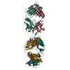
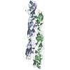
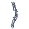
| Title | Loop deletion and proline insertion mutant (deleting six residues and inserted three proline residues) | ||||||
 Components Components | Outer surface protein A | ||||||
 Keywords Keywords | LIPID BINDING PROTEIN / Outer surface protein A / OspA | ||||||
| Function / homology | Outer surface lipoprotein, Borrelia / Outer surface lipoprotein domain superfamily / Borrelia lipoprotein / cell outer membrane / Prokaryotic membrane lipoprotein lipid attachment site profile. / cell surface / membrane / Outer surface protein A Function and homology information Function and homology information | ||||||
| Biological species |  Borrelia burgdorferi (Lyme disease spirochete) Borrelia burgdorferi (Lyme disease spirochete) | ||||||
| Method |  X-RAY DIFFRACTION / SYNCHROTRON / MOLECULAR REPLACEMENT / Resolution: 3 Å X-RAY DIFFRACTION / SYNCHROTRON / MOLECULAR REPLACEMENT / Resolution: 3 Å | ||||||
 Authors Authors | Shiga, S. / Makabe, K. | ||||||
 Citation Citation | Journal: Chembiochem / Year: 2019 Title: Domain-Swapping Design by Polyproline Rod Insertion. Authors: Shiga, S. / Yamanaka, M. / Fujiwara, W. / Hirota, S. / Goda, S. / Makabe, K. | ||||||
| History |
|
- Structure visualization
| Structure viewer | Molecule: MolmilJmol/JSmol |
|---|
- Downloads & links
-Download
| PDBx/mmCIF format | 6iys.cif.gz | 59.6 KB | Display | PDBx/mmCIF format |
|---|---|---|---|---|
| PDB format | pdb6iys.ent.gz | 42 KB | Display | PDB format |
| PDBx/mmJSON format | 6iys.json.gz | Tree view | PDBx/mmJSON format | |
| Others |  Other downloads Other downloads |
-Validation report
| Arichive directory | https://data.pdbj.org/pub/pdb/validation_reports/iy/6iysftp://data.pdbj.org/pub/pdb/validation_reports/iy/6iys | HTTPS FTP |
|---|
-Related structure data
| Related structure data |  6aisC  6icsC  6idcC  6ieiC  2g8cS S: Starting model for refinement C: citing same article ( |
|---|---|
| Similar structure data |


-Links
 PDBj
PDBj


- Assembly
| Deposited unit | 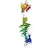
| ||||||||
|---|---|---|---|---|---|---|---|---|---|
| 1 | 
| ||||||||
| Unit cell |
|
-Components
| #1: Protein | Mass: 26154.311 Da / Num. of mol.: 1 Mutation: E37S,E45S,K46S,K48A,K60A,K65S,K83A,E104S,K107S,E196A,K239S,E240S,K254S Source method: isolated from a genetically manipulated source Source: (gene. exp.) Borrelia burgdorferi (strain ATCC 35210 / B31 / CIP 102532 / DSM 4680) (bacteria)Strain: ATCC 35210 / B31 / CIP 102532 / DSM 4680 / Gene: ospA, BB_A15 / Production host: |
|---|---|
| Sequence details | Deletion mutant DSSAAT(205-210) to PPP (205-207) |
-Experimental details
-Experiment
| Experiment | Method: X-RAY DIFFRACTION / Number of used crystals: 1 |
|---|
- Sample preparation
| Crystal | Density Matthews: 4.39 Å3/Da / Density % sol: 71.99 % |
|---|---|
| Crystal grow | Temperature: 292 K / Method: vapor diffusion, hanging drop / pH: 4.7 / Details: 2.5 M Ammonium sulfate 0.1 M sodium acetate |
-Data collection
| Diffraction | Mean temperature: 100 K / Serial crystal experiment: N |
|---|---|
| Diffraction source | Source: SYNCHROTRON / Site: Photon Factory  / Beamline: AR-NE3A / Wavelength: 1 Å / Beamline: AR-NE3A / Wavelength: 1 Å |
| Detector | Type: DECTRIS PILATUS 2M-F / Detector: PIXEL / Date: May 1, 2017 |
| Radiation | Protocol: SINGLE WAVELENGTH / Monochromatic (M) / Laue (L): M / Scattering type: x-ray |
| Radiation wavelength | Wavelength: 1 Å / Relative weight: 1 |
| Reflection | Resolution: 3→20 Å / Num. obs: 18350 / % possible obs: 98.3 % / Redundancy: 7.6 % / Rmerge(I) obs: 0.114 / Net I/σ(I): 30.9 |
| Reflection shell | Resolution: 3→3.05 Å / Redundancy: 7.5 % / Rmerge(I) obs: 0.6 / Mean I/σ(I) obs: 6.15 / Num. unique obs: 946 / % possible all: 100 |
- Processing
| Software |
| ||||||||||||||||||||||||||||||||||||||||||||||||||||||||||||||||||||||||||||||||||||||||||||||||||||||||||||||||||||||||||||||||||||||||||||||||||||||||||||||||||||||||||||||||||||||
|---|---|---|---|---|---|---|---|---|---|---|---|---|---|---|---|---|---|---|---|---|---|---|---|---|---|---|---|---|---|---|---|---|---|---|---|---|---|---|---|---|---|---|---|---|---|---|---|---|---|---|---|---|---|---|---|---|---|---|---|---|---|---|---|---|---|---|---|---|---|---|---|---|---|---|---|---|---|---|---|---|---|---|---|---|---|---|---|---|---|---|---|---|---|---|---|---|---|---|---|---|---|---|---|---|---|---|---|---|---|---|---|---|---|---|---|---|---|---|---|---|---|---|---|---|---|---|---|---|---|---|---|---|---|---|---|---|---|---|---|---|---|---|---|---|---|---|---|---|---|---|---|---|---|---|---|---|---|---|---|---|---|---|---|---|---|---|---|---|---|---|---|---|---|---|---|---|---|---|---|---|---|---|---|
| Refinement | Method to determine structure: MOLECULAR REPLACEMENT Starting model: 2G8C Resolution: 3→20 Å / Cor.coef. Fo:Fc: 0.939 / Cor.coef. Fo:Fc free: 0.92 / SU B: 23.641 / SU ML: 0.385 / Cross valid method: THROUGHOUT / ESU R: 0.814 / ESU R Free: 0.419 / Details: HYDROGENS HAVE BEEN ADDED IN THE RIDING POSITIONS
| ||||||||||||||||||||||||||||||||||||||||||||||||||||||||||||||||||||||||||||||||||||||||||||||||||||||||||||||||||||||||||||||||||||||||||||||||||||||||||||||||||||||||||||||||||||||
| Solvent computation | Ion probe radii: 0.8 Å / Shrinkage radii: 0.8 Å / VDW probe radii: 1.2 Å | ||||||||||||||||||||||||||||||||||||||||||||||||||||||||||||||||||||||||||||||||||||||||||||||||||||||||||||||||||||||||||||||||||||||||||||||||||||||||||||||||||||||||||||||||||||||
| Displacement parameters | Biso mean: 111.437 Å2
| ||||||||||||||||||||||||||||||||||||||||||||||||||||||||||||||||||||||||||||||||||||||||||||||||||||||||||||||||||||||||||||||||||||||||||||||||||||||||||||||||||||||||||||||||||||||
| Refinement step | Cycle: 1 / Resolution: 3→20 Å
| ||||||||||||||||||||||||||||||||||||||||||||||||||||||||||||||||||||||||||||||||||||||||||||||||||||||||||||||||||||||||||||||||||||||||||||||||||||||||||||||||||||||||||||||||||||||
| Refine LS restraints |
|