 Movie
Movie Controller
[English] 日本語
 Yorodumi
- SASDCM7: Truncated monomeric Cytohesin-3 (Grp1; amino acids 63-399) E161A ... -
+ Open data

- Basic information
| Entry | Database: SASBDB / ID: SASDCM7 |
|---|---|
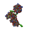
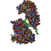
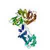
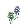
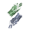
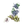
 Sample Sample | Truncated monomeric Cytohesin-3 (Grp1; amino acids 63-399) E161A 6GS Arf6 Q67L fusion protein
|
| Biological species |  |
 Citation Citation | Journal: Structure / Year: 2018 Title: Structural Dynamics Control Allosteric Activation of Cytohesin Family Arf GTPase Exchange Factors. Authors: Andrew W Malaby / Sanchaita Das / Srinivas Chakravarthy / Thomas C Irving / Osman Bilsel / David G Lambright /  Abstract: Membrane dynamic processes including vesicle biogenesis depend on Arf guanosine triphosphatase (GTPase) activation by guanine nucleotide exchange factors (GEFs) containing a catalytic Sec7 domain and ...Membrane dynamic processes including vesicle biogenesis depend on Arf guanosine triphosphatase (GTPase) activation by guanine nucleotide exchange factors (GEFs) containing a catalytic Sec7 domain and a membrane-targeting module such as a pleckstrin homology (PH) domain. The catalytic output of cytohesin family Arf GEFs is controlled by autoinhibitory interactions that impede accessibility of the exchange site in the Sec7 domain. These restraints can be relieved through activator Arf-GTP binding to an allosteric site comprising the PH domain and proximal autoinhibitory elements (Sec7-PH linker and C-terminal helix). Small-angle X-ray scattering and negative-stain electron microscopy were used to investigate the structural organization and conformational dynamics of cytohesin-3 (Grp1) in autoinhibited and active states. The results support a model in which hinge dynamics in the autoinhibited state expose the activator site for Arf-GTP binding, while subsequent C-terminal helix unlatching and repositioning unleash conformational entropy in the Sec7-PH linker to drive exposure of the exchange site. |
 Contact author Contact author |
|
- Structure visualization
| Structure viewer | Molecule:  MolmilJmol/JSmol MolmilJmol/JSmol |
|---|
- Downloads & links
 SASDCM7


-Models
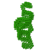
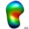
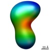
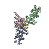
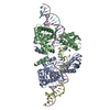
| Model #1556 |  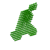 Type: dummy / Software: (r2835) / Radius of dummy atoms: 3.00 A / Symmetry: P1 / Comment: Most representative of 100 dammif models / Chi-square value: 1.050625 / P-value: 0.129700  Search similar-shape structures of this assembly by Omokage search (details) Search similar-shape structures of this assembly by Omokage search (details) |
|---|---|
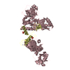
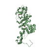
| Model #1557 |  Type: dummy / Radius of dummy atoms: 2.75 A / Symmetry: P1 Comment: Filtered average of 10 filtered averages of 10 dammif models Chi-square value: 1.050625 / P-value: 0.129700 Search similar-shape structures of this assembly by Omokage search (details) |
-Sample
| Sample | Name: Truncated monomeric Cytohesin-3 (Grp1; amino acids 63-399) E161A 6GS Arf6 Q67L fusion protein Specimen concentration: 2.4 mg/ml |
|---|---|
| Buffer | Name: 20 mM Tris, 150 mM NaCl, 2 mM MgCl2, 0.1% 2-mercaptoethanol, 5% glycerol, 0.001 mM insitol 1,3,4,5-tetrakis phosphate pH: 8 |
| Entity #795 | Type: protein / Description: Grp1 63-399 E161A 6GS Arf6 Q67L fusion protein / Formula weight: 61.011 / Num. of mol.: 1 / Source: Mus musculus Sequence: MGHHHHHHGS TTQRNAQIAM GRKKFNMDPK KGIQFLIEND LLQSSPEDVA QFLYKGEGLN KTVIGDYLGE RDDFNIKVLQ AFVELHEFAD LNLVQALRQF LWSFRLPGEA QKIDRMMEAF ASRYCLCNPG VFQSTDTCYV LSFAIIMLNT SLHNHNVRDK PTAERFITMN ...Sequence: MGHHHHHHGS TTQRNAQIAM GRKKFNMDPK KGIQFLIEND LLQSSPEDVA QFLYKGEGLN KTVIGDYLGE RDDFNIKVLQ AFVELHEFAD LNLVQALRQF LWSFRLPGEA QKIDRMMEAF ASRYCLCNPG VFQSTDTCYV LSFAIIMLNT SLHNHNVRDK PTAERFITMN RGINEGGDLP EELLRNLYES IKNEPFKIPE DDGNDLTHTF FNPDREGWLL KLGGRVKTWK RRWFILTDNC LYYFEYTTDK EPRGIIPLEN LSIREVEDPR KPNCFELYNP SHKGQVIKAC KTEADGRVVE GNHVVYRISA PSPEEKEEWM KSIKASISRD PFYDMLATRK RRIANKKGSG SGSGSGSGSG KVLSKIFGNK EMRILMLGLD AAGKTTILYK LKLGQSVTTI PTVGFNVETV TYKNVKFNVW DVGGLDKIRP LWRHYYTGTQ GLIFVVDCAD RDRIDEARQE LHRIINDREM RDAIILIFAN KQDLPDAMKP HEIQEKLGLT RIRDRNWYVQ PSCATSGDGL YEGLTWLTSN YN |
-Experimental information
| Beam | Instrument name: Advanced Photon Source (APS) BioCAT 18ID / City: Argonne, IL / 国: USA / Type of source: X-ray synchrotron / Wavelength: 0.10332 Å / Dist. spec. to detc.: 2.5 mm | ||||||||||||||||||||||||||||||||||||
|---|---|---|---|---|---|---|---|---|---|---|---|---|---|---|---|---|---|---|---|---|---|---|---|---|---|---|---|---|---|---|---|---|---|---|---|---|---|
| Detector | Name: MAR 165 CCD | ||||||||||||||||||||||||||||||||||||
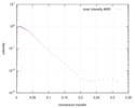
| Scan |  Measurement date: Nov 15, 2013 / Cell temperature: 20 °C / Exposure time: 1 sec. / Number of frames: 1 / Unit: 1/A /
| ||||||||||||||||||||||||||||||||||||
| Distance distribution function P(R) | 
| ||||||||||||||||||||||||||||||||||||
| Result | 
|
