 Movie
Movie Controller

[English] 日本語
 Yorodumi
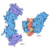
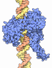
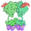
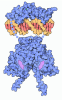
- PDB-9zla: Unbound 106-N-32 nucleosome from SRCAP-CFDP1-nucleosome binding r... -
+ Open data

- Basic information
| Entry | Database: PDB / ID: 9zla | |||||||||||||||||||||||||||||||||
|---|---|---|---|---|---|---|---|---|---|---|---|---|---|---|---|---|---|---|---|---|---|---|---|---|---|---|---|---|---|---|---|---|---|---|
| Title | Unbound 106-N-32 nucleosome from SRCAP-CFDP1-nucleosome binding reaction | |||||||||||||||||||||||||||||||||
 Components Components |
| |||||||||||||||||||||||||||||||||
 Keywords Keywords | GENE REGULATION / Chromatin Remodeler / Snf2 family ATPase / H2A.Z | |||||||||||||||||||||||||||||||||
| Function / homology |  Function and homology information Function and homology informationnucleosomal DNA binding / innate immune response in mucosa / structural constituent of chromatin / nucleosome assembly / nucleosome / chromatin organization / antimicrobial humoral immune response mediated by antimicrobial peptide / heterochromatin formation / antibacterial humoral response / protein heterodimerization activity ...nucleosomal DNA binding / innate immune response in mucosa / structural constituent of chromatin / nucleosome assembly / nucleosome / chromatin organization / antimicrobial humoral immune response mediated by antimicrobial peptide / heterochromatin formation / antibacterial humoral response / protein heterodimerization activity / DNA binding / nucleoplasm / nucleus Similarity search - Function | |||||||||||||||||||||||||||||||||
| Biological species |  Xenopus laevis (African clawed frog) Xenopus laevis (African clawed frog)synthetic construct (others) | |||||||||||||||||||||||||||||||||
| Method | ELECTRON MICROSCOPY / single particle reconstruction / cryo EM / Resolution: 3.1 Å | |||||||||||||||||||||||||||||||||
 Authors Authors | Louder, R.K. / Park, G. | |||||||||||||||||||||||||||||||||
| Funding support |  United States, 1items United States, 1items
| |||||||||||||||||||||||||||||||||
 Citation Citation | Journal: Sci Adv / Year: 2026 Title: Structural mechanism of histone H2A.Z exchange by human SRCAP-CFDP1 holoenzyme. Authors: Giho Park / Carl Wu / Robert K Louder / Abstract: The conserved yeast SWR1 and human SRCAP chromatin remodeling complexes catalyze exchange of nucleosomal histone H2A for H2A.Z, but the underlying mechanism has remained obscure. Here, we show that ...The conserved yeast SWR1 and human SRCAP chromatin remodeling complexes catalyze exchange of nucleosomal histone H2A for H2A.Z, but the underlying mechanism has remained obscure. Here, we show that histone exchange by SRCAP requires the transient activator CFDP1 and resolve nine cryo-electron microscopy structures of the SRCAP-CFDP1 holoenzyme that define the stepwise exchange mechanism. CFDP1 recognizes the conformation of the fully engaged SRCAP-nucleosome complex through interactions with multiple subunits-including direct contact with the ATPase domain-and induces conformational transitions that drive extensive DNA unwrapping, eviction of the H2A-H2B dimer, and insertion of the H2A.Z-H2B dimer, all without necessarily requiring hydrolysis of bound ATP. Collectively, these findings provide unprecedented insight into the mechanism of activator- and nucleotide-driven histone exchange from nucleosomal H2A to H2A.Z. | |||||||||||||||||||||||||||||||||
| History |
|
- Structure visualization
| Structure viewer | Molecule: MolmilJmol/JSmol |
|---|
- Downloads & links
-Download
| PDBx/mmCIF format | 9zla.cif.gz | 348.9 KB | Display | PDBx/mmCIF format |
|---|---|---|---|---|
| PDB format | pdb9zla.ent.gz | 219.1 KB | Display | PDB format |
| PDBx/mmJSON format | 9zla.json.gz | Tree view | PDBx/mmJSON format | |
| Others |  Other downloads Other downloads |
-Validation report
| Arichive directory | https://data.pdbj.org/pub/pdb/validation_reports/zl/9zlaftp://data.pdbj.org/pub/pdb/validation_reports/zl/9zla | HTTPS FTP |
|---|
-Related structure data
| Related structure data |  74396MC  9ca7C  9ca8C  9ca9C  9caaC  9nfvC  9nfwC  9nfxC  9nfyC  9nfzC  9ng0C  9pgcC  9pgdC  9y3dC  9y3eC  9y3fC  9y3gC  9y3hC M: map data used to model this data C: citing same article ( |
|---|---|
| Similar structure data |


-Links
 PDBj
PDBj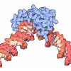
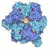

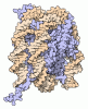


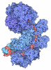
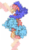
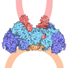

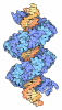
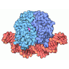






- Assembly
| Deposited unit | 
|
|---|---|
| 1 |
|
-Components
-Protein , 4 types, 8 molecules QSRTUWVX
| #1: Protein | Mass: 13907.163 Da / Num. of mol.: 2 Source method: isolated from a genetically manipulated source Source: (gene. exp.) Xenopus laevis (African clawed frog) / Production host:   Escherichia coli (E. coli) / References: UniProt: P06897 Escherichia coli (E. coli) / References: UniProt: P06897#2: Protein | Mass: 13848.097 Da / Num. of mol.: 2 Source method: isolated from a genetically manipulated source Source: (gene. exp.) Xenopus laevis (African clawed frog) / Production host: Escherichia coli (E. coli) / References: UniProt: P02281#3: Protein | Mass: 15303.930 Da / Num. of mol.: 2 Source method: isolated from a genetically manipulated source Source: (gene. exp.) Xenopus laevis (African clawed frog) / Production host: Escherichia coli (E. coli) / References: UniProt: P84233#4: Protein | Mass: 11263.231 Da / Num. of mol.: 2 Source method: isolated from a genetically manipulated source Source: (gene. exp.) Xenopus laevis (African clawed frog) / Production host: Escherichia coli (E. coli) / References: UniProt: P62799 |
|---|
-DNA chain , 2 types, 2 molecules YZ
| #5: DNA chain | Mass: 87851.664 Da / Num. of mol.: 1 / Source method: obtained synthetically / Source: (synth.) synthetic construct (others) |
|---|---|
| #6: DNA chain | Mass: 88190.930 Da / Num. of mol.: 1 / Source method: obtained synthetically / Source: (synth.) synthetic construct (others) |
-Details
| Has protein modification | N |
|---|
-Experimental details
-Experiment
| Experiment | Method: ELECTRON MICROSCOPY |
|---|---|
| EM experiment | Aggregation state: PARTICLE / 3D reconstruction method: single particle reconstruction |
- Sample preparation
| Component | Name: 106N32 nucleosome / Type: COMPLEX / Entity ID: all / Source: RECOMBINANT | |||||||||
|---|---|---|---|---|---|---|---|---|---|---|
| Molecular weight |
| |||||||||
| Source (natural) | Organism: Xenopus laevis (African clawed frog) | |||||||||
| Source (recombinant) | Organism: Escherichia coli (E. coli) | |||||||||
| Buffer solution | pH: 7.6 | |||||||||
| Specimen | Embedding applied: NO / Shadowing applied: NO / Staining applied: NO / Vitrification applied: YES | |||||||||
| Vitrification | Cryogen name: ETHANE |
- Electron microscopy imaging
| Experimental equipment |  Model: Titan Krios / Image courtesy: FEI Company |
|---|---|
| Microscopy | Model: TFS KRIOS |
| Electron gun | Electron source:  FIELD EMISSION GUN / Accelerating voltage: 300 kV / Illumination mode: FLOOD BEAM FIELD EMISSION GUN / Accelerating voltage: 300 kV / Illumination mode: FLOOD BEAM |
| Electron lens | Mode: BRIGHT FIELD / Nominal defocus max: 1600 nm / Nominal defocus min: 800 nm |
| Image recording | Electron dose: 50 e/Å2 / Film or detector model: GATAN K3 (6k x 4k) |
- Processing
| EM software |
| ||||||||||||||||||||||||
|---|---|---|---|---|---|---|---|---|---|---|---|---|---|---|---|---|---|---|---|---|---|---|---|---|---|
| CTF correction | Type: PHASE FLIPPING AND AMPLITUDE CORRECTION | ||||||||||||||||||||||||
| 3D reconstruction | Resolution: 3.1 Å / Resolution method: FSC 0.143 CUT-OFF / Num. of particles: 93703 / Algorithm: FOURIER SPACE / Symmetry type: POINT | ||||||||||||||||||||||||
| Refinement | Cross valid method: NONE Stereochemistry target values: GeoStd + Monomer Library + CDL v1.2 | ||||||||||||||||||||||||
| Displacement parameters | Biso mean: 47.94 Å2 | ||||||||||||||||||||||||
| Refine LS restraints |
|
