 Movie
Movie Controller

[English] 日本語
 Yorodumi
- EMDB-71626: ARP6-ZNHIT1 module from fully-engaged state of SRCAP-nucleosome c... -
+ Open data

- Basic information
| Entry |  | |||||||||
|---|---|---|---|---|---|---|---|---|---|---|
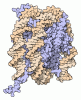
| Title | ARP6-ZNHIT1 module from fully-engaged state of SRCAP-nucleosome complex, with H3-bound ARP6 (focused refinement, filtered by local resolution) | |||||||||
 Map data Map data | ||||||||||
 Sample Sample |
| |||||||||
 Keywords Keywords | Chromatin Remodeler / Snf2 family ATPase / H2A.Z / GENE REGULATION | |||||||||
| Function / homology |  Function and homology information Function and homology informationpositive regulation of lymphoid progenitor cell differentiation / hematopoietic stem cell homeostasis / intestinal stem cell homeostasis / muscle cell differentiation / Swr1 complex / heart process / negative regulation of transcription by RNA polymerase I / nucleolus organization / positive regulation of transcription by RNA polymerase I / positive regulation of DNA damage response, signal transduction by p53 class mediator ...positive regulation of lymphoid progenitor cell differentiation / hematopoietic stem cell homeostasis / intestinal stem cell homeostasis / muscle cell differentiation / Swr1 complex / heart process / negative regulation of transcription by RNA polymerase I / nucleolus organization / positive regulation of transcription by RNA polymerase I / positive regulation of DNA damage response, signal transduction by p53 class mediator / positive regulation of transcription initiation by RNA polymerase II / calcium ion homeostasis / nucleosome binding / transcription initiation-coupled chromatin remodeling / histone deacetylase binding / nucleosomal DNA binding / histone binding / structural constituent of chromatin / nucleosome / heterochromatin formation / cytoskeleton / chromatin remodeling / protein heterodimerization activity / regulation of DNA-templated transcription / nucleolus / nucleoplasm / nucleus Similarity search - Function | |||||||||
| Biological species |  Homo sapiens (human) / Homo sapiens (human) /  Xenopus laevis (African clawed frog) Xenopus laevis (African clawed frog) | |||||||||
| Method | single particle reconstruction / cryo EM / Resolution: 3.2 Å | |||||||||
 Authors Authors | Louder RK / Park G | |||||||||
| Funding support |  United States, 1 items United States, 1 items
| |||||||||
 Citation Citation | Journal: Sci Adv / Year: 2026 Title: Structural mechanism of histone H2A.Z exchange by human SRCAP-CFDP1 holoenzyme. Authors: Giho Park / Carl Wu / Robert K Louder / Abstract: The conserved yeast SWR1 and human SRCAP chromatin remodeling complexes catalyze exchange of nucleosomal histone H2A for H2A.Z, but the underlying mechanism has remained obscure. Here, we show that ...The conserved yeast SWR1 and human SRCAP chromatin remodeling complexes catalyze exchange of nucleosomal histone H2A for H2A.Z, but the underlying mechanism has remained obscure. Here, we show that histone exchange by SRCAP requires the transient activator CFDP1 and resolve nine cryo-electron microscopy structures of the SRCAP-CFDP1 holoenzyme that define the stepwise exchange mechanism. CFDP1 recognizes the conformation of the fully engaged SRCAP-nucleosome complex through interactions with multiple subunits-including direct contact with the ATPase domain-and induces conformational transitions that drive extensive DNA unwrapping, eviction of the H2A-H2B dimer, and insertion of the H2A.Z-H2B dimer, all without necessarily requiring hydrolysis of bound ATP. Collectively, these findings provide unprecedented insight into the mechanism of activator- and nucleotide-driven histone exchange from nucleosomal H2A to H2A.Z. | |||||||||
| History |
|
- Structure visualization
| Supplemental images |
|---|

- Downloads & links
-EMDB archive
| Map data | emd_71626.map.gz | 1.4 MB | EMDB map data format | |
|---|---|---|---|---|
| Header (meta data) | emd-71626-v30.xmlemd-71626.xml | 21.6 KB 21.6 KB | Display Display | EMDB header |
| FSC (resolution estimation) | emd_71626_fsc.xml | 13.6 KB | Display | FSC data file |
| Images |  emd_71626.png emd_71626.png | 102.6 KB | ||
| Masks | emd_71626_msk_1.map | 216 MB | Mask map | |
| Filedesc metadata | emd-71626.cif.gz | 6.7 KB | ||
| Others | emd_71626_half_map_1.map.gzemd_71626_half_map_2.map.gz | 170.6 MB 170.6 MB | ||
| Archive directory |  https://data.pdbj.org/pub/emdb/structures/EMD-71626ftp://data.pdbj.org/pub/emdb/structures/EMD-71626 https://data.pdbj.org/pub/emdb/structures/EMD-71626ftp://data.pdbj.org/pub/emdb/structures/EMD-71626 | HTTPS FTP |
-Related structure data
| Related structure data |  9pgdMC  9ca7C  9ca8C  9ca9C  9caaC  9nfvC  9nfwC  9nfxC  9nfyC  9nfzC  9ng0C  9pgcC  9y3dC  9y3eC  9y3fC  9y3gC  9y3hC  9zlaC M: atomic model generated by this map C: citing same article ( |
|---|---|
| Similar structure data |


-Links
| EMDB pages | EMDB (EBI/PDBe) / EMDataResource |
|---|---|
| Related items in Molecule of the Month |



-Map
| File | Download / File: emd_71626.map.gz / Format: CCP4 / Size: 216 MB / Type: IMAGE STORED AS FLOATING POINT NUMBER (4 BYTES) | ||||||||||||||||||||||||||||||||||||
|---|---|---|---|---|---|---|---|---|---|---|---|---|---|---|---|---|---|---|---|---|---|---|---|---|---|---|---|---|---|---|---|---|---|---|---|---|---|
| Projections & slices | Image control
Images are generated by Spider. | ||||||||||||||||||||||||||||||||||||
| Voxel size | X=Y=Z: 1.025 Å | ||||||||||||||||||||||||||||||||||||
| Density |
| ||||||||||||||||||||||||||||||||||||
| Symmetry | Space group: 1 | ||||||||||||||||||||||||||||||||||||
| Details | EMDB XML:
|
 Z (Sec.)
 Y (Row.)
 X (Col.)


-Supplemental data
-Mask #1
| File | emd_71626_msk_1.map | ||||||||||||
|---|---|---|---|---|---|---|---|---|---|---|---|---|---|
| Projections & Slices |
| ||||||||||||
| Density Histograms |


-Half map: #1
| File | emd_71626_half_map_1.map | ||||||||||||
|---|---|---|---|---|---|---|---|---|---|---|---|---|---|
| Projections & Slices |
| ||||||||||||
| Density Histograms |


-Half map: #2
| File | emd_71626_half_map_2.map | ||||||||||||
|---|---|---|---|---|---|---|---|---|---|---|---|---|---|
| Projections & Slices |
| ||||||||||||
| Density Histograms |


- Sample components
-Entire : SRCAP-nucleosome complex
| Entire | Name: SRCAP-nucleosome complex |
|---|---|
| Components |
|
-Supramolecule #1: SRCAP-nucleosome complex
| Supramolecule | Name: SRCAP-nucleosome complex / type: complex / ID: 1 / Parent: 0 / Macromolecule list: #1-#3 Details: Endogenous purified SRCAP bound to 106N32 nucleosome |
|---|---|
| Source (natural) | Organism: Homo sapiens (human) / Strain: K-562 / Organ: BLOOD / Tissue: BONE MARROW / Organelle: NUCLEUS / Location in cell: NUCLEOPLASM |
-Macromolecule #1: Actin-related protein 6
| Macromolecule | Name: Actin-related protein 6 / type: protein_or_peptide / ID: 1 / Number of copies: 1 / Enantiomer: LEVO |
|---|---|
| Source (natural) | Organism: Homo sapiens (human) / Organ: BLOOD / Tissue: BONE MARROW |
| Molecular weight | Theoretical: 45.857902 KDa |
| Sequence | String: MTTLVLDNGA YNAKIGYSHE NVSVIPNCQF RSKTARLKTF TANQIDEIKD PSGLFYILPF QKGYLVNWDV QRQVWDYLFG KEMYQVDFL DTNIIITEPY FNFTSIQESM NEILFEEYQF QAVLRVNAGA LSAHRYFRDN PSELCCIIVD SGYSFTHIVP Y CRSKKKKE ...String: MTTLVLDNGA YNAKIGYSHE NVSVIPNCQF RSKTARLKTF TANQIDEIKD PSGLFYILPF QKGYLVNWDV QRQVWDYLFG KEMYQVDFL DTNIIITEPY FNFTSIQESM NEILFEEYQF QAVLRVNAGA LSAHRYFRDN PSELCCIIVD SGYSFTHIVP Y CRSKKKKE AIIRINVGGK LLTNHLKEII SYRQLHVMDE THVINQVKED VCYVSQDFYR DMDIAKLKGE ENTVMIDYVL PD FSTIKKG FCKPREEMVL SGKYKSGEQI LRLANERFAV PEILFNPSDI GIQEMGIPEA IVYSIQNLPE EMQPHFFKNI VLT GGNSLF PGFRDRVYSE VRCLTPTDYD VSVVLPENPI TYAWEGGKLI SENDDFEDMV VTREDYEENG HSVCEEKFDI UniProtKB: Actin-related protein 6 |
-Macromolecule #2: Zinc finger HIT domain-containing protein 1
| Macromolecule | Name: Zinc finger HIT domain-containing protein 1 / type: protein_or_peptide / ID: 2 / Number of copies: 1 / Enantiomer: LEVO |
|---|---|
| Source (natural) | Organism: Homo sapiens (human) / Organ: BLOOD / Tissue: BONE MARROW |
| Molecular weight | Theoretical: 17.567023 KDa |
| Sequence | String: MVEKKTSVRS QDPGQRRVLD RAARQRRINR QLEALENDNF QDDPHAGLPQ LGKRLPQFDD DADTGKKKKK TRGDHFKLRF RKNFQALLE EQNLSVAEGP NYLTACAGPP SRPQRPFCAV CGFPSPYTCV SCGARYCTVR CLGTHQETRC LKWTV UniProtKB: Zinc finger HIT domain-containing protein 1 |
-Macromolecule #3: Histone H3.2
| Macromolecule | Name: Histone H3.2 / type: protein_or_peptide / ID: 3 / Number of copies: 1 / Enantiomer: LEVO |
|---|---|
| Source (natural) | Organism: Xenopus laevis (African clawed frog) |
| Molecular weight | Theoretical: 15.30393 KDa |
| Recombinant expression | Organism:   Escherichia coli (E. coli) Escherichia coli (E. coli) |
| Sequence | String: ARTKQTARKS TGGKAPRKQL ATKAARKSAP ATGGVKKPHR YRPGTVALRE IRRYQKSTEL LIRKLPFQRL VREIAQDFKT DLRFQSSAV MALQEASEAY LVALFEDTNL CAIHAKRVTI MPKDIQLARR IRGERA UniProtKB: Histone H3.2 |
-Macromolecule #4: PHOSPHOTHIOPHOSPHORIC ACID-ADENYLATE ESTER
| Macromolecule | Name: PHOSPHOTHIOPHOSPHORIC ACID-ADENYLATE ESTER / type: ligand / ID: 4 / Number of copies: 1 / Formula: AGS |
|---|---|
| Molecular weight | Theoretical: 523.247 Da |
| Chemical component information | 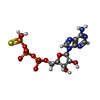 ChemComp-AGS: |
-Macromolecule #5: MAGNESIUM ION
| Macromolecule | Name: MAGNESIUM ION / type: ligand / ID: 5 / Number of copies: 1 / Formula: MG |
|---|---|
| Molecular weight | Theoretical: 24.305 Da |
-Macromolecule #6: ZINC ION
| Macromolecule | Name: ZINC ION / type: ligand / ID: 6 / Number of copies: 2 / Formula: ZN |
|---|---|
| Molecular weight | Theoretical: 65.409 Da |
-Experimental details
-Structure determination
| Method | cryo EM |
|---|---|
 Processing Processing | single particle reconstruction |
| Aggregation state | particle |
-Sample preparation
| Buffer | pH: 7.6 |
|---|---|
| Vitrification | Cryogen name: ETHANE |
- Electron microscopy
| Microscope | TFS KRIOS |
|---|---|
| Image recording | Film or detector model: GATAN K3 (6k x 4k) / Average electron dose: 50.0 e/Å2 |
| Electron beam | Acceleration voltage: 300 kV / Electron source:  FIELD EMISSION GUN FIELD EMISSION GUN |
| Electron optics | Illumination mode: FLOOD BEAM / Imaging mode: BRIGHT FIELD / Nominal defocus max: 1.6 µm / Nominal defocus min: 0.8 µm |
| Experimental equipment |  Model: Titan Krios / Image courtesy: FEI Company |

