 Movie
Movie Controller


+ Open data

- Basic information
| Entry | Database: PDB / ID: 9jhp | |||||||||||||||||||||||||||||||||||||||
|---|---|---|---|---|---|---|---|---|---|---|---|---|---|---|---|---|---|---|---|---|---|---|---|---|---|---|---|---|---|---|---|---|---|---|---|---|---|---|---|---|
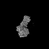
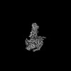
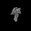
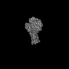
| Title | Cryo-EM structure of GPR4 complexed with miniG13 in pH6.8 | |||||||||||||||||||||||||||||||||||||||
 Components Components |
| |||||||||||||||||||||||||||||||||||||||
 Keywords Keywords | SIGNALING PROTEIN/IMMUNE SYSTEM / GPCR / GPR4 / miniG13 / Proton sensing / SIGNALING PROTEIN / SIGNALING PROTEIN-IMMUNE SYSTEM complex | |||||||||||||||||||||||||||||||||||||||
| Function / homology |  Function and homology information Function and homology informationD5 dopamine receptor binding / regulation of fibroblast migration / Rho-activating G protein-coupled receptor signaling pathway / glomerular mesangial cell development / Class A/1 (Rhodopsin-like receptors) / regulation of vascular permeability / regulation of small GTPase mediated signal transduction / angiogenesis involved in wound healing / response to acidic pH / NRAGE signals death through JNK ...D5 dopamine receptor binding / regulation of fibroblast migration / Rho-activating G protein-coupled receptor signaling pathway / glomerular mesangial cell development / Class A/1 (Rhodopsin-like receptors) / regulation of vascular permeability / regulation of small GTPase mediated signal transduction / angiogenesis involved in wound healing / response to acidic pH / NRAGE signals death through JNK / branching involved in blood vessel morphogenesis / positive regulation of Rho protein signal transduction / CDC42 GTPase cycle / regulation of postsynapse assembly / regulation of cell adhesion / Rho protein signal transduction / cellular response to acidic pH / RAC1 GTPase cycle / guanyl-nucleotide exchange factor activity / negative regulation of angiogenesis / brush border membrane / platelet activation / G protein-coupled receptor activity / regulation of blood pressure / adenylate cyclase-modulating G protein-coupled receptor signaling pathway / G-protein beta/gamma-subunit complex binding / Olfactory Signaling Pathway / Activation of the phototransduction cascade / G protein-coupled acetylcholine receptor signaling pathway / G beta:gamma signalling through PLC beta / Presynaptic function of Kainate receptors / Thromboxane signalling through TP receptor / positive regulation of inflammatory response / Activation of G protein gated Potassium channels / Inhibition of voltage gated Ca2+ channels via Gbeta/gamma subunits / G-protein activation / Glucagon signaling in metabolic regulation / G beta:gamma signalling through CDC42 / Prostacyclin signalling through prostacyclin receptor / G beta:gamma signalling through BTK / Synthesis, secretion, and inactivation of Glucagon-like Peptide-1 (GLP-1) / photoreceptor disc membrane / ADP signalling through P2Y purinoceptor 12 / Sensory perception of sweet, bitter, and umami (glutamate) taste / melanosome / Glucagon-type ligand receptors / Adrenaline,noradrenaline inhibits insulin secretion / Vasopressin regulates renal water homeostasis via Aquaporins / Glucagon-like Peptide-1 (GLP1) regulates insulin secretion / G alpha (z) signalling events / cellular response to catecholamine stimulus / ADP signalling through P2Y purinoceptor 1 / ADORA2B mediated anti-inflammatory cytokines production / G beta:gamma signalling through PI3Kgamma / adenylate cyclase-activating dopamine receptor signaling pathway / regulation of cell shape / Cooperation of PDCL (PhLP1) and TRiC/CCT in G-protein beta folding / GPER1 signaling / cellular response to prostaglandin E stimulus / heterotrimeric G-protein complex / G alpha (12/13) signalling events / Inactivation, recovery and regulation of the phototransduction cascade / G-protein beta-subunit binding / extracellular vesicle / sensory perception of taste / adenylate cyclase-activating G protein-coupled receptor signaling pathway / Thrombin signalling through proteinase activated receptors (PARs) / signaling receptor complex adaptor activity / retina development in camera-type eye / GTPase binding / G protein activity / fibroblast proliferation / Ca2+ pathway / High laminar flow shear stress activates signaling by PIEZO1 and PECAM1:CDH5:KDR in endothelial cells / G alpha (i) signalling events / G alpha (s) signalling events / phospholipase C-activating G protein-coupled receptor signaling pathway / G alpha (q) signalling events / in utero embryonic development / Ras protein signal transduction / cell differentiation / cell population proliferation / Extra-nuclear estrogen signaling / postsynapse / G protein-coupled receptor signaling pathway / lysosomal membrane / focal adhesion / GTPase activity / synapse / GTP binding / protein-containing complex binding / signal transduction / extracellular exosome / membrane / metal ion binding / nucleus / plasma membrane / cytoplasm / cytosol Similarity search - Function | |||||||||||||||||||||||||||||||||||||||
| Biological species |  Homo sapiens (human) Homo sapiens (human) | |||||||||||||||||||||||||||||||||||||||
| Method | ELECTRON MICROSCOPY / single particle reconstruction / cryo EM / Resolution: 3.35 Å | |||||||||||||||||||||||||||||||||||||||
 Authors Authors | Yue, X.L. / Wu, L.J. / Hua, T. / Liu, Z.J. | |||||||||||||||||||||||||||||||||||||||
| Funding support | 1items
| |||||||||||||||||||||||||||||||||||||||
 Citation Citation | Journal: Cell Res / Year: 2025 Title: Structural basis of stepwise proton sensing-mediated GPCR activation. Authors: Xiaolei Yue / Li Peng / Shenhui Liu / Bingjie Zhang / Xiaodan Zhang / Hao Chang / Yuan Pei / Xiaoting Li / Junlin Liu / Wenqing Shui / Lijie Wu / Huji Xu / Zhi-Jie Liu / Tian Hua /  Abstract: The regulation of pH homeostasis is crucial in many biological processes vital for survival, growth, and function of life. The pH-sensing G protein-coupled receptors (GPCRs), including GPR4, GPR65 ...The regulation of pH homeostasis is crucial in many biological processes vital for survival, growth, and function of life. The pH-sensing G protein-coupled receptors (GPCRs), including GPR4, GPR65 and GPR68, play a pivotal role in detecting changes in extracellular proton concentrations, impacting both physiological and pathological states. However, comprehensive understanding of the proton sensing mechanism is still elusive. Here, we determined the cryo-electron microscopy structures of GPR4 and GPR65 in various activation states across different pH levels, coupled with G, G or G proteins, as well as a small molecule NE52-QQ57-bound inactive GPR4 structure. These structures reveal the dynamic nature of the extracellular loop 2 and its signature conformations in different receptor states, and disclose the proton sensing mechanism mediated by networks of extracellular histidine and carboxylic acid residues. Notably, we unexpectedly captured partially active intermediate states of both GPR4-G and GPR4-G complexes, and identified a unique allosteric binding site for NE52-QQ57 in GPR4. By integrating prior investigations with our structural analysis and mutagenesis data, we propose a detailed atomic model for stepwise proton sensation and GPCR activation. These insights may pave the way for the development of selective ligands and targeted therapeutic interventions for pH sensing-relevant diseases. | |||||||||||||||||||||||||||||||||||||||
| History |
|
- Structure visualization
| Structure viewer | Molecule: MolmilJmol/JSmol |
|---|
- Downloads & links
-Download
| PDBx/mmCIF format | 9jhp.cif.gz | 234.4 KB | Display | PDBx/mmCIF format |
|---|---|---|---|---|
| PDB format | pdb9jhp.ent.gz | 180.1 KB | Display | PDB format |
| PDBx/mmJSON format | 9jhp.json.gz | Tree view | PDBx/mmJSON format | |
| Others |  Other downloads Other downloads |
-Validation report
| Arichive directory | https://data.pdbj.org/pub/pdb/validation_reports/jh/9jhpftp://data.pdbj.org/pub/pdb/validation_reports/jh/9jhp | HTTPS FTP |
|---|
-Related structure data
| Related structure data | 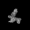 61489MC 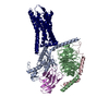 8zceC 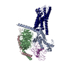 8zcfC  9jftC  9jfuC 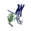 9jfvC  9jfwC 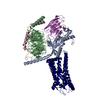 9jfxC 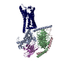 9jfzC 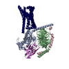 9lgmC M: map data used to model this data C: citing same article ( |
|---|---|
| Similar structure data |


-Links
 PDBj
PDBj


- Assembly
| Deposited unit | 
|
|---|---|
| 1 |
|
-Components
| #1: Protein | Mass: 26574.400 Da / Num. of mol.: 1 Source method: isolated from a genetically manipulated source Source: (gene. exp.) Homo sapiens (human) / Gene: GNA13 / Production host:   Spodoptera frugiperda (fall armyworm) / References: UniProt: Q14344 Spodoptera frugiperda (fall armyworm) / References: UniProt: Q14344 |
|---|---|
| #2: Protein | Mass: 38045.629 Da / Num. of mol.: 1 Source method: isolated from a genetically manipulated source Source: (gene. exp.) Homo sapiens (human) / Gene: GNB1 / Production host: Spodoptera frugiperda (fall armyworm) / References: UniProt: P62873 |
| #3: Antibody | Mass: 30668.211 Da / Num. of mol.: 1 Source method: isolated from a genetically manipulated source Source: (gene. exp.) Homo sapiens (human) / Production host: Spodoptera frugiperda (fall armyworm) |
| #4: Protein | Mass: 7861.143 Da / Num. of mol.: 1 Source method: isolated from a genetically manipulated source Source: (gene. exp.) Homo sapiens (human) / Gene: GNG2 / Production host: Spodoptera frugiperda (fall armyworm) / References: UniProt: P59768 |
| #5: Protein | Mass: 42688.301 Da / Num. of mol.: 1 Source method: isolated from a genetically manipulated source Source: (gene. exp.) Homo sapiens (human) / Gene: GPR4 / Production host: Spodoptera frugiperda (fall armyworm) / References: UniProt: P46093 |
| Has protein modification | Y |
-Experimental details
-Experiment
| Experiment | Method: ELECTRON MICROSCOPY |
|---|---|
| EM experiment | Aggregation state: PARTICLE / 3D reconstruction method: single particle reconstruction |
- Sample preparation
| Component | Name: gpr4-g13 / Type: COMPLEX / Entity ID: all / Source: RECOMBINANT |
|---|---|
| Source (natural) | Organism: Homo sapiens (human) |
| Source (recombinant) | Organism: Spodoptera frugiperda (fall armyworm) |
| Buffer solution | pH: 6.8 |
| Specimen | Embedding applied: NO / Shadowing applied: NO / Staining applied: NO / Vitrification applied: YES |
| Vitrification | Cryogen name: ETHANE |
- Electron microscopy imaging
| Experimental equipment |  Model: Talos Arctica / Image courtesy: FEI Company |
|---|---|
| Microscopy | Model: FEI TALOS ARCTICA |
| Electron gun | Electron source:  FIELD EMISSION GUN / Accelerating voltage: 300 kV / Illumination mode: FLOOD BEAM FIELD EMISSION GUN / Accelerating voltage: 300 kV / Illumination mode: FLOOD BEAM |
| Electron lens | Mode: BRIGHT FIELD / Nominal defocus max: 2000 nm / Nominal defocus min: 1200 nm |
| Image recording | Electron dose: 60 e/Å2 / Film or detector model: FEI FALCON IV (4k x 4k) |
- Processing
| EM software | Name: PHENIX / Category: model refinement | ||||||||||||||||||||||||
|---|---|---|---|---|---|---|---|---|---|---|---|---|---|---|---|---|---|---|---|---|---|---|---|---|---|
| CTF correction | Type: PHASE FLIPPING ONLY | ||||||||||||||||||||||||
| 3D reconstruction | Resolution: 3.35 Å / Resolution method: FSC 0.143 CUT-OFF / Num. of particles: 171879 / Symmetry type: POINT | ||||||||||||||||||||||||
| Refinement | Highest resolution: 3.35 Å Stereochemistry target values: REAL-SPACE (WEIGHTED MAP SUM AT ATOM CENTERS) | ||||||||||||||||||||||||
| Refine LS restraints |
|
