 Movie
Movie Controller


+ Open data

- Basic information
| Entry |  | |||||||||
|---|---|---|---|---|---|---|---|---|---|---|
| Title | Cryo-EM structure of GPR4 complexed with miniG13 in pH6.8 | |||||||||
 Map data Map data | ||||||||||
 Sample Sample |
| |||||||||
 Keywords Keywords | GPCR / GPR4 / miniG13 / Proton sensing / SIGNALING PROTEIN / SIGNALING PROTEIN-IMMUNE SYSTEM complex | |||||||||
| Function / homology |  Function and homology information Function and homology informationD5 dopamine receptor binding / regulation of fibroblast migration / Rho-activating G protein-coupled receptor signaling pathway / regulation of vascular permeability / Class A/1 (Rhodopsin-like receptors) / regulation of small GTPase mediated signal transduction / response to acidic pH / NRAGE signals death through JNK / branching involved in blood vessel morphogenesis / positive regulation of Rho protein signal transduction ...D5 dopamine receptor binding / regulation of fibroblast migration / Rho-activating G protein-coupled receptor signaling pathway / regulation of vascular permeability / Class A/1 (Rhodopsin-like receptors) / regulation of small GTPase mediated signal transduction / response to acidic pH / NRAGE signals death through JNK / branching involved in blood vessel morphogenesis / positive regulation of Rho protein signal transduction / CDC42 GTPase cycle / regulation of postsynapse assembly / regulation of cell adhesion / Rho protein signal transduction / cellular response to acidic pH / RAC1 GTPase cycle / guanyl-nucleotide exchange factor activity / brush border membrane / platelet activation / regulation of blood pressure / G protein-coupled receptor activity / G-protein beta/gamma-subunit complex binding / adenylate cyclase-modulating G protein-coupled receptor signaling pathway / Olfactory Signaling Pathway / Activation of the phototransduction cascade / positive regulation of inflammatory response / G protein-coupled acetylcholine receptor signaling pathway / G beta:gamma signalling through PLC beta / Presynaptic function of Kainate receptors / Thromboxane signalling through TP receptor / Activation of G protein gated Potassium channels / Inhibition of voltage gated Ca2+ channels via Gbeta/gamma subunits / G-protein activation / Glucagon signaling in metabolic regulation / Prostacyclin signalling through prostacyclin receptor / G beta:gamma signalling through CDC42 / melanosome / Synthesis, secretion, and inactivation of Glucagon-like Peptide-1 (GLP-1) / G beta:gamma signalling through BTK / photoreceptor disc membrane / ADP signalling through P2Y purinoceptor 12 / Glucagon-type ligand receptors / Sensory perception of sweet, bitter, and umami (glutamate) taste / Adrenaline,noradrenaline inhibits insulin secretion / Vasopressin regulates renal water homeostasis via Aquaporins / Glucagon-like Peptide-1 (GLP1) regulates insulin secretion / G alpha (z) signalling events / regulation of cell shape / cellular response to catecholamine stimulus / ADP signalling through P2Y purinoceptor 1 / ADORA2B mediated anti-inflammatory cytokines production / G beta:gamma signalling through PI3Kgamma / adenylate cyclase-activating dopamine receptor signaling pathway / Cooperation of PDCL (PhLP1) and TRiC/CCT in G-protein beta folding / GPER1 signaling / cellular response to prostaglandin E stimulus / heterotrimeric G-protein complex / G alpha (12/13) signalling events / Inactivation, recovery and regulation of the phototransduction cascade / G-protein beta-subunit binding / extracellular vesicle / sensory perception of taste / Thrombin signalling through proteinase activated receptors (PARs) / adenylate cyclase-activating G protein-coupled receptor signaling pathway / signaling receptor complex adaptor activity / retina development in camera-type eye / GTPase binding / fibroblast proliferation / G protein activity / Ca2+ pathway / High laminar flow shear stress activates signaling by PIEZO1 and PECAM1:CDH5:KDR in endothelial cells / G alpha (i) signalling events / in utero embryonic development / G alpha (s) signalling events / G alpha (q) signalling events / phospholipase C-activating G protein-coupled receptor signaling pathway / Ras protein signal transduction / cell differentiation / Extra-nuclear estrogen signaling / cell population proliferation / postsynapse / G protein-coupled receptor signaling pathway / lysosomal membrane / focal adhesion / GTPase activity / synapse / GTP binding / protein-containing complex binding / signal transduction / extracellular exosome / membrane / metal ion binding / nucleus / plasma membrane / cytoplasm / cytosol Similarity search - Function | |||||||||
| Biological species |  Homo sapiens (human) Homo sapiens (human) | |||||||||
| Method | single particle reconstruction / cryo EM / Resolution: 3.35 Å | |||||||||
 Authors Authors | Yue XL / Wu LJ / Hua T / Liu ZJ | |||||||||
| Funding support | 1 items
| |||||||||
 Citation Citation | Journal: Cell Res / Year: 2025 Title: Structural basis of stepwise proton sensing-mediated GPCR activation. Authors: Xiaolei Yue / Li Peng / Shenhui Liu / Bingjie Zhang / Xiaodan Zhang / Hao Chang / Yuan Pei / Xiaoting Li / Junlin Liu / Wenqing Shui / Lijie Wu / Huji Xu / Zhi-Jie Liu / Tian Hua /  Abstract: The regulation of pH homeostasis is crucial in many biological processes vital for survival, growth, and function of life. The pH-sensing G protein-coupled receptors (GPCRs), including GPR4, GPR65 ...The regulation of pH homeostasis is crucial in many biological processes vital for survival, growth, and function of life. The pH-sensing G protein-coupled receptors (GPCRs), including GPR4, GPR65 and GPR68, play a pivotal role in detecting changes in extracellular proton concentrations, impacting both physiological and pathological states. However, comprehensive understanding of the proton sensing mechanism is still elusive. Here, we determined the cryo-electron microscopy structures of GPR4 and GPR65 in various activation states across different pH levels, coupled with G, G or G proteins, as well as a small molecule NE52-QQ57-bound inactive GPR4 structure. These structures reveal the dynamic nature of the extracellular loop 2 and its signature conformations in different receptor states, and disclose the proton sensing mechanism mediated by networks of extracellular histidine and carboxylic acid residues. Notably, we unexpectedly captured partially active intermediate states of both GPR4-G and GPR4-G complexes, and identified a unique allosteric binding site for NE52-QQ57 in GPR4. By integrating prior investigations with our structural analysis and mutagenesis data, we propose a detailed atomic model for stepwise proton sensation and GPCR activation. These insights may pave the way for the development of selective ligands and targeted therapeutic interventions for pH sensing-relevant diseases. | |||||||||
| History |
|
- Structure visualization
| Supplemental images |
|---|

- Downloads & links
-EMDB archive
| Map data | emd_61489.map.gz | 56.6 MB | EMDB map data format | |
|---|---|---|---|---|
| Header (meta data) | emd-61489-v30.xmlemd-61489.xml | 21 KB 21 KB | Display Display | EMDB header |
| FSC (resolution estimation) | emd_61489_fsc.xml | 8.4 KB | Display | FSC data file |
| Images |  emd_61489.png emd_61489.png | 59.3 KB | ||
| Filedesc metadata | emd-61489.cif.gz | 6.8 KB | ||
| Others | emd_61489_half_map_1.map.gzemd_61489_half_map_2.map.gz | 59.5 MB 59.5 MB | ||
| Archive directory |  http://ftp.pdbj.org/pub/emdb/structures/EMD-61489ftp://ftp.pdbj.org/pub/emdb/structures/EMD-61489 http://ftp.pdbj.org/pub/emdb/structures/EMD-61489ftp://ftp.pdbj.org/pub/emdb/structures/EMD-61489 | HTTPS FTP |
-Related structure data
| Related structure data | 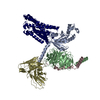 9jhpMC 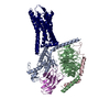 8zceC 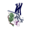 8zcfC  9jftC 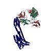 9jfuC 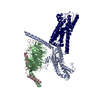 9jfvC  9jfwC 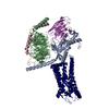 9jfxC 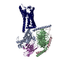 9jfzC 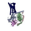 9lgmC M: atomic model generated by this map C: citing same article ( |
|---|---|
| Similar structure data |


-Links
| EMDB pages | EMDB (EBI/PDBe) / EMDataResource |
|---|---|
| Related items in Molecule of the Month |


-Map
| File | Download / File: emd_61489.map.gz / Format: CCP4 / Size: 64 MB / Type: IMAGE STORED AS FLOATING POINT NUMBER (4 BYTES) | ||||||||||||||||||||||||||||||||||||
|---|---|---|---|---|---|---|---|---|---|---|---|---|---|---|---|---|---|---|---|---|---|---|---|---|---|---|---|---|---|---|---|---|---|---|---|---|---|
| Projections & slices | Image control
Images are generated by Spider. | ||||||||||||||||||||||||||||||||||||
| Voxel size | X=Y=Z: 0.96 Å | ||||||||||||||||||||||||||||||||||||
| Density |
| ||||||||||||||||||||||||||||||||||||
| Symmetry | Space group: 1 | ||||||||||||||||||||||||||||||||||||
| Details | EMDB XML:
|
 Z (Sec.)
 Y (Row.)
 X (Col.)


-Supplemental data
-Half map: #2
| File | emd_61489_half_map_1.map | ||||||||||||
|---|---|---|---|---|---|---|---|---|---|---|---|---|---|
| Projections & Slices |
| ||||||||||||
| Density Histograms |


-Half map: #1
| File | emd_61489_half_map_2.map | ||||||||||||
|---|---|---|---|---|---|---|---|---|---|---|---|---|---|
| Projections & Slices |
| ||||||||||||
| Density Histograms |


- Sample components
-Entire : gpr4-g13
| Entire | Name: gpr4-g13 |
|---|---|
| Components |
|
-Supramolecule #1: gpr4-g13
| Supramolecule | Name: gpr4-g13 / type: complex / ID: 1 / Parent: 0 / Macromolecule list: all |
|---|---|
| Source (natural) | Organism: Homo sapiens (human) |
-Macromolecule #1: Guanine nucleotide-binding protein subunit alpha-13
| Macromolecule | Name: Guanine nucleotide-binding protein subunit alpha-13 / type: protein_or_peptide / ID: 1 / Number of copies: 1 / Enantiomer: LEVO |
|---|---|
| Source (natural) | Organism: Homo sapiens (human) |
| Molecular weight | Theoretical: 26.5744 KDa |
| Recombinant expression | Organism:   Spodoptera frugiperda (fall armyworm) Spodoptera frugiperda (fall armyworm) |
| Sequence | String: MGSTVSAEDK AAAERSKEID KCLSREKTYV KRLVKILLLG ADNSGKSTFL KQMRIIHGGS GGSGGTKGIH EYDFEIKNVP FKMVDVGGQ RSERKRWFEC FDSVTSILFL VDSSDFNRLT ESLNDFETIV NNRVFSNVSI ILFLNKTDLL EEKVQIVSIK D YFLEFEGD ...String: MGSTVSAEDK AAAERSKEID KCLSREKTYV KRLVKILLLG ADNSGKSTFL KQMRIIHGGS GGSGGTKGIH EYDFEIKNVP FKMVDVGGQ RSERKRWFEC FDSVTSILFL VDSSDFNRLT ESLNDFETIV NNRVFSNVSI ILFLNKTDLL EEKVQIVSIK D YFLEFEGD PHCLRDVQKF LVECFRNKRR DQQQKPLYHH FTTAINTENA RLIFRDVKDT ILHDNLKQLM LQ UniProtKB: Guanine nucleotide-binding protein subunit alpha-13, Guanine nucleotide-binding protein subunit alpha-13 |
-Macromolecule #2: Guanine nucleotide-binding protein G(I)/G(S)/G(T) subunit beta-1
| Macromolecule | Name: Guanine nucleotide-binding protein G(I)/G(S)/G(T) subunit beta-1 type: protein_or_peptide / ID: 2 / Number of copies: 1 / Enantiomer: LEVO |
|---|---|
| Source (natural) | Organism: Homo sapiens (human) |
| Molecular weight | Theoretical: 38.045629 KDa |
| Recombinant expression | Organism: Spodoptera frugiperda (fall armyworm) |
| Sequence | String: IGRARGFSEL DQLRQEAEQL KNQIRDARKA CADATLSQIT NNIDPVGRIQ MRTRRTLRGH LAKIYAMHWG TDSRLLVSAS QDGKLIIWD SYTTNKVHAI PLRSSWVMTC AYAPSGNYVA CGGLDNICSI YNLKTREGNV RVSRELAGHT GYLSCCRFLD D NQIVTSSG ...String: IGRARGFSEL DQLRQEAEQL KNQIRDARKA CADATLSQIT NNIDPVGRIQ MRTRRTLRGH LAKIYAMHWG TDSRLLVSAS QDGKLIIWD SYTTNKVHAI PLRSSWVMTC AYAPSGNYVA CGGLDNICSI YNLKTREGNV RVSRELAGHT GYLSCCRFLD D NQIVTSSG DTTCALWDIE TGQQTTTFTG HTGDVMSLSL APDTRLFVSG ACDASAKLWD VREGMCRQTF TGHESDINAI CF FPNGNAF ATGSDDATCR LFDLRADQEL MTYSHDNIIC GITSVSFSKS GRLLLAGYDD FNCNVWDALK ADRAGVLAGH DNR VSCLGV TDDGMAVATG SWDSFLKIWN UniProtKB: Guanine nucleotide-binding protein G(I)/G(S)/G(T) subunit beta-1 |
-Macromolecule #3: scFv16
| Macromolecule | Name: scFv16 / type: protein_or_peptide / ID: 3 / Number of copies: 1 / Enantiomer: LEVO |
|---|---|
| Source (natural) | Organism: Homo sapiens (human) |
| Molecular weight | Theoretical: 30.668211 KDa |
| Recombinant expression | Organism: Spodoptera frugiperda (fall armyworm) |
| Sequence | String: MVSAIVLYVL LAAAAHSAFA DVQLVESGGG LVQPGGSRKL SCSASGFAFS SFGMHWVRQA PEKGLEWVAY ISSGSGTIYY ADTVKGRFT ISRDDPKNTL FLQMTSLRSE DTAMYYCVRS IYYYGSSPFD FWGQGTTLTV SSGGGGSGGG GSGGGGSDIV M TQATSSVP ...String: MVSAIVLYVL LAAAAHSAFA DVQLVESGGG LVQPGGSRKL SCSASGFAFS SFGMHWVRQA PEKGLEWVAY ISSGSGTIYY ADTVKGRFT ISRDDPKNTL FLQMTSLRSE DTAMYYCVRS IYYYGSSPFD FWGQGTTLTV SSGGGGSGGG GSGGGGSDIV M TQATSSVP VTPGESVSIS CRSSKSLLHS NGNTYLYWFL QRPGQSPQLL IYRMSNLASG VPDRFSGSGS GTAFTLTISR LE AEDVGVY YCMQHLEYPL TFGAGTKLEL KAAAENLYFQ SHHHHHHHH |
-Macromolecule #4: Guanine nucleotide-binding protein G(I)/G(S)/G(O) subunit gamma-2
| Macromolecule | Name: Guanine nucleotide-binding protein G(I)/G(S)/G(O) subunit gamma-2 type: protein_or_peptide / ID: 4 / Number of copies: 1 / Enantiomer: LEVO |
|---|---|
| Source (natural) | Organism: Homo sapiens (human) |
| Molecular weight | Theoretical: 7.861143 KDa |
| Recombinant expression | Organism: Spodoptera frugiperda (fall armyworm) |
| Sequence | String: MASNNTASIA QARKLVEQLK MEANIDRIKV SKAAADLMAY CEAHAKEDPL LTPVPASENP FREKKFFCAI L UniProtKB: Guanine nucleotide-binding protein G(I)/G(S)/G(O) subunit gamma-2 |
-Macromolecule #5: G-protein coupled receptor 4
| Macromolecule | Name: G-protein coupled receptor 4 / type: protein_or_peptide / ID: 5 / Number of copies: 1 / Enantiomer: LEVO |
|---|---|
| Source (natural) | Organism: Homo sapiens (human) |
| Molecular weight | Theoretical: 42.688301 KDa |
| Recombinant expression | Organism: Spodoptera frugiperda (fall armyworm) |
| Sequence | String: MGNHTWEGCH VDSRVDHLFP PSLYIFVIGV GLPTNCLALW AAYRQVQQRN ELGVYLMNLS IADLLYICTL PLWVDYFLHH DNWIHGPGS CKLFGFIFYT NIYISIAFLC CISVDRYLAV AHPLRFARLR RVKTAVAVSS VVWATELGAN SAPLFHDELF R DRYNHTFC ...String: MGNHTWEGCH VDSRVDHLFP PSLYIFVIGV GLPTNCLALW AAYRQVQQRN ELGVYLMNLS IADLLYICTL PLWVDYFLHH DNWIHGPGS CKLFGFIFYT NIYISIAFLC CISVDRYLAV AHPLRFARLR RVKTAVAVSS VVWATELGAN SAPLFHDELF R DRYNHTFC FEKFPMEGWV AWMNLYRVFV GFLFPWALML LSYRGILRAV RGSVSTERQE KAKIKRLALS LIAIVLVCFA PY HVLLLSR SAIYLGRPWD CGFEERVFSA YHSSLAFTSL NCVADPILYC LVNEGARSDV AKALHNLLRF LASDKPQEMA NAS LTLETP LTSKRNSTAK AMTGSWAATP PSQGDQVQEF LEVLFQGPHH HHHHHHHH UniProtKB: G protein-coupled receptor 4 |
-Experimental details
-Structure determination
| Method | cryo EM |
|---|---|
 Processing Processing | single particle reconstruction |
| Aggregation state | particle |
-Sample preparation
| Buffer | pH: 6.8 |
|---|---|
| Vitrification | Cryogen name: ETHANE |
- Electron microscopy
| Microscope | FEI TALOS ARCTICA |
|---|---|
| Image recording | Film or detector model: FEI FALCON IV (4k x 4k) / Average electron dose: 60.0 e/Å2 |
| Electron beam | Acceleration voltage: 300 kV / Electron source:  FIELD EMISSION GUN FIELD EMISSION GUN |
| Electron optics | Illumination mode: FLOOD BEAM / Imaging mode: BRIGHT FIELD / Nominal defocus max: 2.0 µm / Nominal defocus min: 1.2 µm |
| Experimental equipment |  Model: Talos Arctica / Image courtesy: FEI Company |

