 Movie
Movie Controller


+ Open data

- Basic information
| Entry | Database: PDB / ID: 9jfu | |||||||||||||||||||||||||||||||||||||||
|---|---|---|---|---|---|---|---|---|---|---|---|---|---|---|---|---|---|---|---|---|---|---|---|---|---|---|---|---|---|---|---|---|---|---|---|---|---|---|---|---|
| Title | Cryo-EM structure of inactive GPR4 with NE52-QQ57 | |||||||||||||||||||||||||||||||||||||||
 Components Components |
| |||||||||||||||||||||||||||||||||||||||
 Keywords Keywords | SIGNALING PROTEIN/IMMUNE SYSTEM / GPCR / Gs / Gsq / Proton sensing / SIGNALING PROTEIN / SIGNALING PROTEIN-IMMUNE SYSTEM complex | |||||||||||||||||||||||||||||||||||||||
| Function / homology |  Function and homology information Function and homology informationregulation of vascular permeability / Class A/1 (Rhodopsin-like receptors) / response to acidic pH / positive regulation of Rho protein signal transduction / regulation of cell adhesion / cellular response to acidic pH / electron transport chain / G protein-coupled receptor activity / positive regulation of inflammatory response / phospholipase C-activating G protein-coupled receptor signaling pathway ...regulation of vascular permeability / Class A/1 (Rhodopsin-like receptors) / response to acidic pH / positive regulation of Rho protein signal transduction / regulation of cell adhesion / cellular response to acidic pH / electron transport chain / G protein-coupled receptor activity / positive regulation of inflammatory response / phospholipase C-activating G protein-coupled receptor signaling pathway / adenylate cyclase-activating G protein-coupled receptor signaling pathway / G alpha (q) signalling events / electron transfer activity / periplasmic space / iron ion binding / G protein-coupled receptor signaling pathway / heme binding / plasma membrane Similarity search - Function | |||||||||||||||||||||||||||||||||||||||
| Biological species |  Homo sapiens (human) Homo sapiens (human) | |||||||||||||||||||||||||||||||||||||||
| Method | ELECTRON MICROSCOPY / single particle reconstruction / cryo EM / Resolution: 3.23 Å | |||||||||||||||||||||||||||||||||||||||
 Authors Authors | Yue, X.L. / Wu, L.J. / Hua, T. / Liu, Z.J. | |||||||||||||||||||||||||||||||||||||||
| Funding support | 1items
| |||||||||||||||||||||||||||||||||||||||
 Citation Citation | Journal: Cell Res / Year: 2025 Title: Structural basis of stepwise proton sensing-mediated GPCR activation. Authors: Xiaolei Yue / Li Peng / Shenhui Liu / Bingjie Zhang / Xiaodan Zhang / Hao Chang / Yuan Pei / Xiaoting Li / Junlin Liu / Wenqing Shui / Lijie Wu / Huji Xu / Zhi-Jie Liu / Tian Hua /  Abstract: The regulation of pH homeostasis is crucial in many biological processes vital for survival, growth, and function of life. The pH-sensing G protein-coupled receptors (GPCRs), including GPR4, GPR65 ...The regulation of pH homeostasis is crucial in many biological processes vital for survival, growth, and function of life. The pH-sensing G protein-coupled receptors (GPCRs), including GPR4, GPR65 and GPR68, play a pivotal role in detecting changes in extracellular proton concentrations, impacting both physiological and pathological states. However, comprehensive understanding of the proton sensing mechanism is still elusive. Here, we determined the cryo-electron microscopy structures of GPR4 and GPR65 in various activation states across different pH levels, coupled with G, G or G proteins, as well as a small molecule NE52-QQ57-bound inactive GPR4 structure. These structures reveal the dynamic nature of the extracellular loop 2 and its signature conformations in different receptor states, and disclose the proton sensing mechanism mediated by networks of extracellular histidine and carboxylic acid residues. Notably, we unexpectedly captured partially active intermediate states of both GPR4-G and GPR4-G complexes, and identified a unique allosteric binding site for NE52-QQ57 in GPR4. By integrating prior investigations with our structural analysis and mutagenesis data, we propose a detailed atomic model for stepwise proton sensation and GPCR activation. These insights may pave the way for the development of selective ligands and targeted therapeutic interventions for pH sensing-relevant diseases. | |||||||||||||||||||||||||||||||||||||||
| History |
|
- Structure visualization
| Structure viewer | Molecule: MolmilJmol/JSmol |
|---|
- Downloads & links
-Download
| PDBx/mmCIF format | 9jfu.cif.gz | 203.5 KB | Display | PDBx/mmCIF format |
|---|---|---|---|---|
| PDB format | pdb9jfu.ent.gz | Display | PDB format | |
| PDBx/mmJSON format | 9jfu.json.gz | Tree view | PDBx/mmJSON format | |
| Others |  Other downloads Other downloads |
-Validation report
| Arichive directory | https://data.pdbj.org/pub/pdb/validation_reports/jf/9jfuftp://data.pdbj.org/pub/pdb/validation_reports/jf/9jfu | HTTPS FTP |
|---|
-Related structure data
| Related structure data |  61440MC 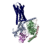 8zceC 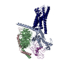 8zcfC  9jftC 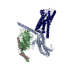 9jfvC  9jfwC  9jfxC  9jfzC  9jhpC  9lgmC M: map data used to model this data C: citing same article ( |
|---|---|
| Similar structure data |


-Links
 PDBj
PDBj


- Assembly
| Deposited unit | 
|
|---|---|
| 1 |
|
-Components
| #1: Antibody | Mass: 66624.789 Da / Num. of mol.: 1 Source method: isolated from a genetically manipulated source Source: (gene. exp.) Homo sapiens (human) / Cell line (production host): HEK293 / Production host: Homo sapiens (human) |
|---|---|
| #2: Antibody | Mass: 13390.644 Da / Num. of mol.: 1 Source method: isolated from a genetically manipulated source Source: (gene. exp.) Homo sapiens (human) / Production host:  |
| #3: Antibody | Mass: 23353.947 Da / Num. of mol.: 1 Source method: isolated from a genetically manipulated source Source: (gene. exp.) Homo sapiens (human) / Cell line (production host): HEK293 / Production host: Homo sapiens (human) |
| #4: Protein | Mass: 55236.461 Da / Num. of mol.: 1 Source method: isolated from a genetically manipulated source Source: (gene. exp.) Homo sapiens (human) / Gene: GPR4, cybC / Production host:   Spodoptera frugiperda (fall armyworm) / References: UniProt: P46093, UniProt: P0ABE7 Spodoptera frugiperda (fall armyworm) / References: UniProt: P46093, UniProt: P0ABE7 |
| #5: Chemical | ChemComp-A1L1E / Mass: 416.519 Da / Num. of mol.: 1 / Source method: obtained synthetically / Formula: C24H28N6O / Feature type: SUBJECT OF INVESTIGATION |
| Has ligand of interest | Y |
| Has protein modification | Y |
-Experimental details
-Experiment
| Experiment | Method: ELECTRON MICROSCOPY |
|---|---|
| EM experiment | Aggregation state: PARTICLE / 3D reconstruction method: single particle reconstruction |
- Sample preparation
| Component |
| ||||||||||||||||||||||||
|---|---|---|---|---|---|---|---|---|---|---|---|---|---|---|---|---|---|---|---|---|---|---|---|---|---|
| Source (natural) |
| ||||||||||||||||||||||||
| Source (recombinant) | Organism: Spodoptera frugiperda (fall armyworm) | ||||||||||||||||||||||||
| Buffer solution | pH: 7.5 | ||||||||||||||||||||||||
| Specimen | Embedding applied: NO / Shadowing applied: NO / Staining applied: NO / Vitrification applied: YES | ||||||||||||||||||||||||
| Vitrification | Cryogen name: ETHANE |
- Electron microscopy imaging
| Experimental equipment |  Model: Talos Arctica / Image courtesy: FEI Company |
|---|---|
| Microscopy | Model: FEI TALOS ARCTICA |
| Electron gun | Electron source:  FIELD EMISSION GUN / Accelerating voltage: 300 kV / Illumination mode: FLOOD BEAM FIELD EMISSION GUN / Accelerating voltage: 300 kV / Illumination mode: FLOOD BEAM |
| Electron lens | Mode: BRIGHT FIELD / Nominal defocus max: 2000 nm / Nominal defocus min: 1200 nm |
| Image recording | Electron dose: 60 e/Å2 / Film or detector model: GATAN K3 BIOCONTINUUM (6k x 4k) |
- Processing
| EM software | Name: PHENIX / Category: model refinement | ||||||||||||||||||||||||
|---|---|---|---|---|---|---|---|---|---|---|---|---|---|---|---|---|---|---|---|---|---|---|---|---|---|
| CTF correction | Type: PHASE FLIPPING ONLY | ||||||||||||||||||||||||
| 3D reconstruction | Resolution: 3.23 Å / Resolution method: FSC 0.143 CUT-OFF / Num. of particles: 151171 / Symmetry type: POINT | ||||||||||||||||||||||||
| Refinement | Highest resolution: 3.23 Å Stereochemistry target values: REAL-SPACE (WEIGHTED MAP SUM AT ATOM CENTERS) | ||||||||||||||||||||||||
| Refine LS restraints |
|
