 Movie
Movie Controller

[English] 日本語
 Yorodumi
- PDB-9j25: Structural basis of the bifunctionality of M. salinexigens ZYF650... -
+ Open data

- Basic information
| Entry | Database: PDB / ID: 9j25 | ||||||
|---|---|---|---|---|---|---|---|
| Title | Structural basis of the bifunctionality of M. salinexigens ZYF650T glucosylglycerol phosphorylase in glucosylglycerol catabolism | ||||||
 Components Components | Sucrose phosphorylase | ||||||
 Keywords Keywords | STRUCTURAL PROTEIN / Glucosylglycerol / phosphorylase-GH13_18 / structure-double displacement mechanism | ||||||
| Function / homology |  Function and homology information Function and homology informationsucrose phosphorylase / sucrose phosphorylase activity / carbohydrate metabolic process Similarity search - Function | ||||||
| Biological species |  Marinobacter salinexigens (bacteria) Marinobacter salinexigens (bacteria) | ||||||
| Method |  X-RAY DIFFRACTION / SYNCHROTRON / MOLECULAR REPLACEMENT / Resolution: 2.74 Å X-RAY DIFFRACTION / SYNCHROTRON / MOLECULAR REPLACEMENT / Resolution: 2.74 Å | ||||||
 Authors Authors | Lu, D. / Ma, H.L. | ||||||
| Funding support |  China, 1items China, 1items
| ||||||
 Citation Citation | Journal: J Biol Chem / Year: 2025 Title: Structural basis of the bifunctionality of Marinobacter salinexigens ZYF650 glucosylglycerol phosphorylase in glucosylglycerol catabolism. Authors: Di Lu / Keke Zhang / Chen Cheng / Danni Wu / Lei Yin / Quan Luo / Meiyun Shi / Honglei Ma / Xuefeng Lu / Abstract: 2-O-α-Glucosylglycerol (GG) is a natural heteroside synthesized by many cyanobacteria and a few heterotrophic bacteria under salt stress conditions. Bacteria produce GG in response to stimuli and ...2-O-α-Glucosylglycerol (GG) is a natural heteroside synthesized by many cyanobacteria and a few heterotrophic bacteria under salt stress conditions. Bacteria produce GG in response to stimuli and degrade it once the stimulus diminishes. Heterotrophic bacteria utilize GG phosphorylase (GGP), a member of the GH13_18 family, via a two-step process consisting of phosphorolysis and hydrolysis for GG catabolism. However, the precise mechanism by which GGP degrades GG remains elusive. We determined the 3D structure of a recently identified GGP (MsGGP) of the deep-sea bacterium Marinobacter salinexigens ZYF650, in complex with glucose and glycerol, α-d-glucose-1-phosphate (αGlc1-P), and orthophosphate (inorganic phosphate) at resolutions of 2.5, 2.7, and 2.7 Å, respectively. Notably, the first αGlc1-P complex structure in the GH13_18 family, the complex of MsGGP and αGlc1-P, validates that GGP catalyzes GG decomposition through consecutive phosphorolysis and hydrolysis. In addition, the structure reveals the mechanism of high stereoselectivity on αGlc1-P. Glu231 and Asp190 were identified as the catalytic residues. Interestingly, these structures closely resemble each other, indicating minimal conformational changes upon binding end-product glucose and glycerol, or the intermediate αGlc1-P. The structures also indicate that the substrates may follow a specific trajectory and a precise order toward the active center in close proximity and in a geometrically favorable orientation for catalysis in a double displacement mechanism. | ||||||
| History |
|
- Structure visualization
| Structure viewer | Molecule: MolmilJmol/JSmol |
|---|
- Downloads & links
-Download
| PDBx/mmCIF format | 9j25.cif.gz | 536.3 KB | Display | PDBx/mmCIF format |
|---|---|---|---|---|
| PDB format | pdb9j25.ent.gz | 447.3 KB | Display | PDB format |
| PDBx/mmJSON format | 9j25.json.gz | Tree view | PDBx/mmJSON format | |
| Others |  Other downloads Other downloads |
-Validation report
| Arichive directory | https://data.pdbj.org/pub/pdb/validation_reports/j2/9j25ftp://data.pdbj.org/pub/pdb/validation_reports/j2/9j25 | HTTPS FTP |
|---|
-Related structure data
| Related structure data |  9j1uC  9j22C  9j24C 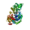 7xdqS S: Starting model for refinement C: citing same article ( |
|---|---|
| Similar structure data |

-Links
 PDBj
PDBj

- Assembly
| Deposited unit | 
| |||||||||
|---|---|---|---|---|---|---|---|---|---|---|
| 1 | 
| |||||||||
| Unit cell |
| |||||||||
| Components on special symmetry positions |
|
-Components
-Protein , 1 types, 3 molecules ABC
| #1: Protein | Mass: 54903.852 Da / Num. of mol.: 3 Source method: isolated from a genetically manipulated source Source: (gene. exp.) Marinobacter salinexigens (bacteria) / Gene: gtfA, FWJ25_14990 / Production host: |
|---|
-Non-polymers , 5 types, 169 molecules 


| #2: Chemical | ChemComp-PEG /  Mass: 106.120 Da / Num. of mol.: 1 / Source method: obtained synthetically / Formula: C4H10O3 Mass: 106.120 Da / Num. of mol.: 1 / Source method: obtained synthetically / Formula: C4H10O3 | ||||
|---|---|---|---|---|---|
| #3: Chemical | ChemComp-TRS /  Mass: 122.143 Da / Num. of mol.: 1 / Source method: obtained synthetically / Formula: C4H12NO3 / Comment: pH buffer*YM Mass: 122.143 Da / Num. of mol.: 1 / Source method: obtained synthetically / Formula: C4H12NO3 / Comment: pH buffer*YM | ||||
| #4: Chemical |  Mass: 94.971 Da / Num. of mol.: 3 / Source method: obtained synthetically / Formula: PO4 / Feature type: SUBJECT OF INVESTIGATION Mass: 94.971 Da / Num. of mol.: 3 / Source method: obtained synthetically / Formula: PO4 / Feature type: SUBJECT OF INVESTIGATION#5: Chemical |  Mass: 96.063 Da / Num. of mol.: 2 / Source method: obtained synthetically / Formula: SO4 Mass: 96.063 Da / Num. of mol.: 2 / Source method: obtained synthetically / Formula: SO4#6: Water | ChemComp-HOH / | Mass: 18.015 Da / Num. of mol.: 162 / Source method: isolated from a natural source / Formula: H2O |
-Details
| Has ligand of interest | Y |
|---|---|
| Has protein modification | N |
-Experimental details
-Experiment
| Experiment | Method: X-RAY DIFFRACTION / Number of used crystals: 1 |
|---|
- Sample preparation
| Crystal | Density Matthews: 2.54 Å3/Da / Density % sol: 51.62 % |
|---|---|
| Crystal grow | Temperature: 289 K / Method: vapor diffusion Details: 1.8 M Ammonium sulfate, 0.1 M BIS-TRIS pH 6.5, 2% Polyethylene glycol monomethyl ether 550 |
-Data collection
| Diffraction | Mean temperature: 100 K / Serial crystal experiment: N |
|---|---|
| Diffraction source | Source: SYNCHROTRON / Site: SSRF / Beamline: BL18U1 / Wavelength: 0.987 Å |
| Detector | Type: DECTRIS PILATUS 6M / Detector: PIXEL / Date: Dec 17, 2023 |
| Radiation | Protocol: SINGLE WAVELENGTH / Monochromatic (M) / Laue (L): M / Scattering type: x-ray |
| Radiation wavelength | Wavelength: 0.987 Å / Relative weight: 1 |
| Reflection | Resolution: 2.74→50 Å / Num. obs: 44128 / % possible obs: 90.47 % / Redundancy: 12.3 % / R split: 0.055 / Rrim(I) all: 0.196 / Net I/σ(I): 8.4 |
| Reflection shell | Resolution: 2.75→2.8 Å / Num. unique obs: 2165 / Rpim(I) all: 0.348 |
- Processing
| Software |
| ||||||||||||||||||||||||||||||||||||||||||||||||||||||||||||||||||||||||||||||||||||||||||||||||||||||||||||||||||||||||||||||||||||||||||||||||||||||||||||||||||||||||||||||||||||||||||||||||||||||||||||||||||||||||||||||||||||||||||||||||||||||||||||||||||||||||||||||||||||||||||||||||||||||||||||
|---|---|---|---|---|---|---|---|---|---|---|---|---|---|---|---|---|---|---|---|---|---|---|---|---|---|---|---|---|---|---|---|---|---|---|---|---|---|---|---|---|---|---|---|---|---|---|---|---|---|---|---|---|---|---|---|---|---|---|---|---|---|---|---|---|---|---|---|---|---|---|---|---|---|---|---|---|---|---|---|---|---|---|---|---|---|---|---|---|---|---|---|---|---|---|---|---|---|---|---|---|---|---|---|---|---|---|---|---|---|---|---|---|---|---|---|---|---|---|---|---|---|---|---|---|---|---|---|---|---|---|---|---|---|---|---|---|---|---|---|---|---|---|---|---|---|---|---|---|---|---|---|---|---|---|---|---|---|---|---|---|---|---|---|---|---|---|---|---|---|---|---|---|---|---|---|---|---|---|---|---|---|---|---|---|---|---|---|---|---|---|---|---|---|---|---|---|---|---|---|---|---|---|---|---|---|---|---|---|---|---|---|---|---|---|---|---|---|---|---|---|---|---|---|---|---|---|---|---|---|---|---|---|---|---|---|---|---|---|---|---|---|---|---|---|---|---|---|---|---|---|---|---|---|---|---|---|---|---|---|---|---|---|---|---|---|---|---|---|---|---|---|---|---|---|---|---|---|---|---|---|---|---|---|---|---|---|---|---|---|---|---|---|---|---|---|---|---|---|---|---|---|
| Refinement | Method to determine structure: MOLECULAR REPLACEMENT Starting model: 7XDQ Resolution: 2.74→49.09 Å / SU ML: 0.38 / Cross valid method: FREE R-VALUE / σ(F): 1.35 / Phase error: 28.09 / Stereochemistry target values: ML
| ||||||||||||||||||||||||||||||||||||||||||||||||||||||||||||||||||||||||||||||||||||||||||||||||||||||||||||||||||||||||||||||||||||||||||||||||||||||||||||||||||||||||||||||||||||||||||||||||||||||||||||||||||||||||||||||||||||||||||||||||||||||||||||||||||||||||||||||||||||||||||||||||||||||||||||
| Solvent computation | Shrinkage radii: 0.9 Å / VDW probe radii: 1.11 Å / Solvent model: FLAT BULK SOLVENT MODEL | ||||||||||||||||||||||||||||||||||||||||||||||||||||||||||||||||||||||||||||||||||||||||||||||||||||||||||||||||||||||||||||||||||||||||||||||||||||||||||||||||||||||||||||||||||||||||||||||||||||||||||||||||||||||||||||||||||||||||||||||||||||||||||||||||||||||||||||||||||||||||||||||||||||||||||||
| Refinement step | Cycle: LAST / Resolution: 2.74→49.09 Å
| ||||||||||||||||||||||||||||||||||||||||||||||||||||||||||||||||||||||||||||||||||||||||||||||||||||||||||||||||||||||||||||||||||||||||||||||||||||||||||||||||||||||||||||||||||||||||||||||||||||||||||||||||||||||||||||||||||||||||||||||||||||||||||||||||||||||||||||||||||||||||||||||||||||||||||||
| Refine LS restraints |
| ||||||||||||||||||||||||||||||||||||||||||||||||||||||||||||||||||||||||||||||||||||||||||||||||||||||||||||||||||||||||||||||||||||||||||||||||||||||||||||||||||||||||||||||||||||||||||||||||||||||||||||||||||||||||||||||||||||||||||||||||||||||||||||||||||||||||||||||||||||||||||||||||||||||||||||
| LS refinement shell |
| ||||||||||||||||||||||||||||||||||||||||||||||||||||||||||||||||||||||||||||||||||||||||||||||||||||||||||||||||||||||||||||||||||||||||||||||||||||||||||||||||||||||||||||||||||||||||||||||||||||||||||||||||||||||||||||||||||||||||||||||||||||||||||||||||||||||||||||||||||||||||||||||||||||||||||||
| Refinement TLS params. | Method: refined / Refine-ID: X-RAY DIFFRACTION
| ||||||||||||||||||||||||||||||||||||||||||||||||||||||||||||||||||||||||||||||||||||||||||||||||||||||||||||||||||||||||||||||||||||||||||||||||||||||||||||||||||||||||||||||||||||||||||||||||||||||||||||||||||||||||||||||||||||||||||||||||||||||||||||||||||||||||||||||||||||||||||||||||||||||||||||
| Refinement TLS group |
|
