+ Open data
- Basic information
| Entry |  | |||||||||
|---|---|---|---|---|---|---|---|---|---|---|
| Title | structure of human urea transport protein slc14A1 with urea | |||||||||
 Map data Map data | ||||||||||
 Sample Sample |
| |||||||||
 Keywords Keywords | human urea transport protein slc14A1 / TRANSPORT PROTEIN | |||||||||
| Function / homology |  Function and homology information Function and homology informationurea channel activity / water transmembrane transporter activity / urea transport / : / urea transmembrane transporter activity / urea transmembrane transport / establishment of localization in cell / transmembrane transport / basolateral plasma membrane / plasma membrane Similarity search - Function | |||||||||
| Biological species |  Homo sapiens (human) Homo sapiens (human) | |||||||||
| Method | single particle reconstruction / cryo EM / Resolution: 2.75 Å | |||||||||
 Authors Authors | He J / Wang F / Zhong C / Zhang P / Liu Z | |||||||||
| Funding support |  China, 1 items China, 1 items
| |||||||||
 Citation Citation |  Journal: J Biol Chem / Year: 2025 Journal: J Biol Chem / Year: 2025Title: Structural basis of the bifunctionality of Marinobacter salinexigens ZYF650 glucosylglycerol phosphorylase in glucosylglycerol catabolism. Authors: Di Lu / Keke Zhang / Chen Cheng / Danni Wu / Lei Yin / Quan Luo / Meiyun Shi / Honglei Ma / Xuefeng Lu /  Abstract: 2-O-α-Glucosylglycerol (GG) is a natural heteroside synthesized by many cyanobacteria and a few heterotrophic bacteria under salt stress conditions. Bacteria produce GG in response to stimuli and ...2-O-α-Glucosylglycerol (GG) is a natural heteroside synthesized by many cyanobacteria and a few heterotrophic bacteria under salt stress conditions. Bacteria produce GG in response to stimuli and degrade it once the stimulus diminishes. Heterotrophic bacteria utilize GG phosphorylase (GGP), a member of the GH13_18 family, via a two-step process consisting of phosphorolysis and hydrolysis for GG catabolism. However, the precise mechanism by which GGP degrades GG remains elusive. We determined the 3D structure of a recently identified GGP (MsGGP) of the deep-sea bacterium Marinobacter salinexigens ZYF650, in complex with glucose and glycerol, α-d-glucose-1-phosphate (αGlc1-P), and orthophosphate (inorganic phosphate) at resolutions of 2.5, 2.7, and 2.7 Å, respectively. Notably, the first αGlc1-P complex structure in the GH13_18 family, the complex of MsGGP and αGlc1-P, validates that GGP catalyzes GG decomposition through consecutive phosphorolysis and hydrolysis. In addition, the structure reveals the mechanism of high stereoselectivity on αGlc1-P. Glu231 and Asp190 were identified as the catalytic residues. Interestingly, these structures closely resemble each other, indicating minimal conformational changes upon binding end-product glucose and glycerol, or the intermediate αGlc1-P. The structures also indicate that the substrates may follow a specific trajectory and a precise order toward the active center in close proximity and in a geometrically favorable orientation for catalysis in a double displacement mechanism. | |||||||||
| History |
|
- Structure visualization
| Supplemental images |
|---|
- Downloads & links
-EMDB archive
| Map data |  emd_61086.map.gz emd_61086.map.gz | 59.8 MB |  EMDB map data format EMDB map data format | |
|---|---|---|---|---|
| Header (meta data) |  emd-61086-v30.xml emd-61086-v30.xml emd-61086.xml emd-61086.xml | 15.6 KB 15.6 KB | Display Display |  EMDB header EMDB header |
| Images |  emd_61086.png emd_61086.png | 119 KB | ||
| Filedesc metadata |  emd-61086.cif.gz emd-61086.cif.gz | 5.6 KB | ||
| Others |  emd_61086_half_map_1.map.gz emd_61086_half_map_1.map.gz emd_61086_half_map_2.map.gz emd_61086_half_map_2.map.gz | 59.3 MB 59.3 MB | ||
| Archive directory |  http://ftp.pdbj.org/pub/emdb/structures/EMD-61086 http://ftp.pdbj.org/pub/emdb/structures/EMD-61086 ftp://ftp.pdbj.org/pub/emdb/structures/EMD-61086 ftp://ftp.pdbj.org/pub/emdb/structures/EMD-61086 | HTTPS FTP |
-Validation report
| Summary document |  emd_61086_validation.pdf.gz emd_61086_validation.pdf.gz | 866.5 KB | Display |  EMDB validaton report EMDB validaton report |
|---|---|---|---|---|
| Full document |  emd_61086_full_validation.pdf.gz emd_61086_full_validation.pdf.gz | 866.1 KB | Display | |
| Data in XML |  emd_61086_validation.xml.gz emd_61086_validation.xml.gz | 12.4 KB | Display | |
| Data in CIF |  emd_61086_validation.cif.gz emd_61086_validation.cif.gz | 14.5 KB | Display | |
| Arichive directory |  https://ftp.pdbj.org/pub/emdb/validation_reports/EMD-61086 https://ftp.pdbj.org/pub/emdb/validation_reports/EMD-61086 ftp://ftp.pdbj.org/pub/emdb/validation_reports/EMD-61086 ftp://ftp.pdbj.org/pub/emdb/validation_reports/EMD-61086 | HTTPS FTP |
-Related structure data
| Related structure data |  9j22MC  9j1uC  9j24C  9j25C M: atomic model generated by this map C: citing same article ( |
|---|---|
| Similar structure data | Similarity search - Function & homology  F&H Search F&H Search |
- Links
Links
| EMDB pages |  EMDB (EBI/PDBe) / EMDB (EBI/PDBe) /  EMDataResource EMDataResource |
|---|
- Map
Map
| File |  Download / File: emd_61086.map.gz / Format: CCP4 / Size: 64 MB / Type: IMAGE STORED AS FLOATING POINT NUMBER (4 BYTES) Download / File: emd_61086.map.gz / Format: CCP4 / Size: 64 MB / Type: IMAGE STORED AS FLOATING POINT NUMBER (4 BYTES) | ||||||||||||||||||||||||||||||||||||
|---|---|---|---|---|---|---|---|---|---|---|---|---|---|---|---|---|---|---|---|---|---|---|---|---|---|---|---|---|---|---|---|---|---|---|---|---|---|
| Projections & slices | Image control
Images are generated by Spider. | ||||||||||||||||||||||||||||||||||||
| Voxel size | X=Y=Z: 0.83 Å | ||||||||||||||||||||||||||||||||||||
| Density |
| ||||||||||||||||||||||||||||||||||||
| Symmetry | Space group: 1 | ||||||||||||||||||||||||||||||||||||
| Details | EMDB XML:
|
-Supplemental data
-Half map: #2
| File | emd_61086_half_map_1.map | ||||||||||||
|---|---|---|---|---|---|---|---|---|---|---|---|---|---|
| Projections & Slices |
| ||||||||||||
| Density Histograms |
-Half map: #1
| File | emd_61086_half_map_2.map | ||||||||||||
|---|---|---|---|---|---|---|---|---|---|---|---|---|---|
| Projections & Slices |
| ||||||||||||
| Density Histograms |
- Sample components
-Entire : Complex of human urea transport protein slc14A1 with urea
| Entire | Name: Complex of human urea transport protein slc14A1 with urea |
|---|---|
| Components |
|
-Supramolecule #1: Complex of human urea transport protein slc14A1 with urea
| Supramolecule | Name: Complex of human urea transport protein slc14A1 with urea type: organelle_or_cellular_component / ID: 1 / Parent: 0 / Macromolecule list: #1 |
|---|---|
| Source (natural) | Organism:  Homo sapiens (human) Homo sapiens (human) |
-Macromolecule #1: Urea transporter 1
| Macromolecule | Name: Urea transporter 1 / type: protein_or_peptide / ID: 1 / Number of copies: 3 / Enantiomer: LEVO |
|---|---|
| Source (natural) | Organism:  Homo sapiens (human) Homo sapiens (human) |
| Molecular weight | Theoretical: 42.566145 KDa |
| Recombinant expression | Organism:  Baculoviridae sp. (virus) Baculoviridae sp. (virus) |
| Sequence | String: MEDSPTMVRV DSPTMVRGEN QVSPCQGRRC FPKALGYVTG DMKELANQLK DKPVVLQFID WILRGISQVV FVNNPVSGIL ILVGLLVQN PWWALTGWLG TVVSTLMALL LSQDRSLIAS GLYGYNATLV GVLMAVFSDK GDYFWWLLLP VCAMSMTCPI F SSALNSML ...String: MEDSPTMVRV DSPTMVRGEN QVSPCQGRRC FPKALGYVTG DMKELANQLK DKPVVLQFID WILRGISQVV FVNNPVSGIL ILVGLLVQN PWWALTGWLG TVVSTLMALL LSQDRSLIAS GLYGYNATLV GVLMAVFSDK GDYFWWLLLP VCAMSMTCPI F SSALNSML SKWDLPVFTL PFNMALSMYL SATGHYNPFF PAKLVIPITT APQISWSDLS ALELLKSIPV GVGQIYGCDN PW TGGIFLG AILLSSPLMC LHAAIGSLLG IAAGLSLSAP FEDIYFGLWG FNSSLACIAM GGMFMALTWQ THLLALGCAL FTA YLGVGM ANFMAEVGLP ACTWPFCLAT LLFLIMTTKN SNIYKMPLSK VTYPEENRIF YLQAKKRMVE SPL UniProtKB: Urea transporter 1 |
-Macromolecule #2: UREA
| Macromolecule | Name: UREA / type: ligand / ID: 2 / Number of copies: 15 / Formula: URE |
|---|---|
| Molecular weight | Theoretical: 60.055 Da |
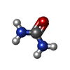
| Chemical component information |  ChemComp-URE: |
-Macromolecule #3: PALMITIC ACID
| Macromolecule | Name: PALMITIC ACID / type: ligand / ID: 3 / Number of copies: 12 / Formula: PLM |
|---|---|
| Molecular weight | Theoretical: 256.424 Da |
| Chemical component information |  ChemComp-PLM: |
-Experimental details
-Structure determination
| Method | cryo EM |
|---|---|
 Processing Processing | single particle reconstruction |
| Aggregation state | particle |
- Sample preparation
| Buffer | pH: 7.4 |
|---|---|
| Sugar embedding | Material: water |
| Grid | Model: Quantifoil R1.2/1.3 / Pretreatment - Type: PLASMA CLEANING |
| Vitrification | Cryogen name: ETHANE |
- Electron microscopy
| Microscope | FEI TITAN KRIOS |
|---|---|
| Image recording | Film or detector model: GATAN K3 BIOQUANTUM (6k x 4k) / Average electron dose: 80.0 e/Å2 |
| Electron beam | Acceleration voltage: 300 kV / Electron source:  FIELD EMISSION GUN FIELD EMISSION GUN |
| Electron optics | Illumination mode: FLOOD BEAM / Imaging mode: BRIGHT FIELD / Nominal defocus max: 1.5 µm / Nominal defocus min: 1.0 µm |
| Experimental equipment |  Model: Titan Krios / Image courtesy: FEI Company |
 Movie
Movie Controller


 Z (Sec.)
 Y (Row.)
 X (Col.)


