 Movie
Movie Controller

[English] 日本語
 Yorodumi
- PDB-7xdq: Crystal structure of a glucosylglycerol phosphorylase mutant from... -
+ Open data

- Basic information
| Entry | Database: PDB / ID: 7xdq | ||||||
|---|---|---|---|---|---|---|---|
| Title | Crystal structure of a glucosylglycerol phosphorylase mutant from Marinobacter adhaerens | ||||||
 Components Components | Glucosylglycerol phosphorylase | ||||||
 Keywords Keywords | BIOSYNTHETIC PROTEIN / phosphorylase | ||||||
| Function / homology |  Function and homology information Function and homology informationglucosylglycerol phosphorylase (configuration-retaining) / glucosylglycerol metabolic process / 1,4-alpha-oligoglucan phosphorylase activity / glycosyltransferase activity Similarity search - Function | ||||||
| Biological species |  Marinobacter adhaerens (bacteria) Marinobacter adhaerens (bacteria) | ||||||
| Method |  X-RAY DIFFRACTION / SYNCHROTRON / MOLECULAR REPLACEMENT / Resolution: 2.83 Å X-RAY DIFFRACTION / SYNCHROTRON / MOLECULAR REPLACEMENT / Resolution: 2.83 Å | ||||||
 Authors Authors | Wei, H.L. / Li, Q. / Yang, J.G. / Liu, W.D. / Sun, Y.X. | ||||||
| Funding support | 1items
| ||||||
 Citation Citation | Journal: Acs Catalysis / Year: 2022 Title: Protein Engineering of Glucosylglycerol Phosphorylase Facilitating Efficient and Highly Regio- and Stereoselective Glycosylation of Polyols in a Synthetic System. Authors: Zhang, T. / Liu, P. / Wei, H. / Sun, X. / Zeng, Y. / Zhang, X. / Cai, Y. / Cui, M. / Ma, H. / Liu, W. / Sun, Y. / Yang, J. | ||||||
| History |
|
- Structure visualization
| Structure viewer | Molecule: MolmilJmol/JSmol |
|---|
- Downloads & links
-Download
| PDBx/mmCIF format | 7xdq.cif.gz | 108.9 KB | Display | PDBx/mmCIF format |
|---|---|---|---|---|
| PDB format | pdb7xdq.ent.gz | 82 KB | Display | PDB format |
| PDBx/mmJSON format | 7xdq.json.gz | Tree view | PDBx/mmJSON format | |
| Others |  Other downloads Other downloads |
-Validation report
| Arichive directory | https://data.pdbj.org/pub/pdb/validation_reports/xd/7xdqftp://data.pdbj.org/pub/pdb/validation_reports/xd/7xdq | HTTPS FTP |
|---|
-Related structure data
| Related structure data | 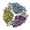 7xdrC  6s9vS S: Starting model for refinement C: citing same article ( |
|---|---|
| Similar structure data |
-Links
 PDBj
PDBj


- Assembly
| Deposited unit | 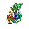
| ||||||||
|---|---|---|---|---|---|---|---|---|---|
| 1 |
| ||||||||
| Unit cell |
|
-Components
| #1: Protein | Mass: 54930.984 Da / Num. of mol.: 1 Source method: isolated from a genetically manipulated source Source: (gene. exp.) Marinobacter adhaerens (bacteria) / Strain: DSM 23420 / HP15 / Gene: gtfA, HP15_2853 / Plasmid: pET-28a / Production host: References: UniProt: E4PMA5, glucosylglycerol phosphorylase (configuration-retaining) |
|---|---|
| #2: Sugar | ChemComp-BGC /   Type: D-saccharide, beta linking / Mass: 180.156 Da / Num. of mol.: 1 / Source method: obtained synthetically / Formula: C6H12O6 / Feature type: SUBJECT OF INVESTIGATION Type: D-saccharide, beta linking / Mass: 180.156 Da / Num. of mol.: 1 / Source method: obtained synthetically / Formula: C6H12O6 / Feature type: SUBJECT OF INVESTIGATION |
| #3: Chemical | ChemComp-LI /   Mass: 6.941 Da / Num. of mol.: 1 / Source method: obtained synthetically / Formula: Li Mass: 6.941 Da / Num. of mol.: 1 / Source method: obtained synthetically / Formula: Li |
| #4: Water | ChemComp-HOH /  Mass: 18.015 Da / Num. of mol.: 1 / Source method: isolated from a natural source / Formula: H2O Mass: 18.015 Da / Num. of mol.: 1 / Source method: isolated from a natural source / Formula: H2O |
| Has ligand of interest | Y |
-Experimental details
-Experiment
| Experiment | Method: X-RAY DIFFRACTION / Number of used crystals: 1 |
|---|
- Sample preparation
| Crystal | Density Matthews: 3.48 Å3/Da / Density % sol: 64.68 % |
|---|---|
| Crystal grow | Temperature: 295 K / Method: vapor diffusion, sitting drop / pH: 8 / Details: PEG8000, MgAc |
-Data collection
| Diffraction | Mean temperature: 100 K / Serial crystal experiment: N |
|---|---|
| Diffraction source | Source: SYNCHROTRON / Site: SSRF  / Beamline: BL02U1 / Wavelength: 1 Å / Beamline: BL02U1 / Wavelength: 1 Å |
| Detector | Type: DECTRIS EIGER X 16M / Detector: PIXEL / Date: Jan 1, 2022 |
| Radiation | Monochromator: GRAPHITE / Protocol: SINGLE WAVELENGTH / Monochromatic (M) / Laue (L): M / Scattering type: x-ray |
| Radiation wavelength | Wavelength: 1 Å / Relative weight: 1 |
| Reflection | Resolution: 2.82→30 Å / Num. obs: 18371 / % possible obs: 100 % / Redundancy: 13.1 % / CC1/2: 1 / Rpim(I) all: 0.031 / Net I/σ(I): 32.03 |
| Reflection shell | Resolution: 2.82→2.92 Å / Num. unique obs: 1800 / CC1/2: 0.746 / Rpim(I) all: 0.435 |
- Processing
| Software |
| ||||||||||||||||||||||||||||||||||||||||||||||||||||||||
|---|---|---|---|---|---|---|---|---|---|---|---|---|---|---|---|---|---|---|---|---|---|---|---|---|---|---|---|---|---|---|---|---|---|---|---|---|---|---|---|---|---|---|---|---|---|---|---|---|---|---|---|---|---|---|---|---|---|
| Refinement | Method to determine structure: MOLECULAR REPLACEMENT Starting model: 6S9V Resolution: 2.83→21.52 Å / SU ML: 0.53 / Cross valid method: THROUGHOUT / σ(F): 1.35 / Phase error: 35.11 / Stereochemistry target values: ML
| ||||||||||||||||||||||||||||||||||||||||||||||||||||||||
| Solvent computation | Shrinkage radii: 0.9 Å / VDW probe radii: 1.1 Å / Solvent model: FLAT BULK SOLVENT MODEL | ||||||||||||||||||||||||||||||||||||||||||||||||||||||||
| Displacement parameters | Biso max: 161.65 Å2 / Biso mean: 92.3111 Å2 / Biso min: 31.24 Å2 | ||||||||||||||||||||||||||||||||||||||||||||||||||||||||
| Refinement step | Cycle: final / Resolution: 2.83→21.52 Å
| ||||||||||||||||||||||||||||||||||||||||||||||||||||||||
| LS refinement shell | Refine-ID: X-RAY DIFFRACTION / Rfactor Rfree error: 0 / Total num. of bins used: 7
|
