 Movie
Movie Controller

[English] 日本語
 Yorodumi
- PDB-8rue: Crystal structure of Rhizobium etli L-asparaginase ReAV H139A mutant -
+ Open data

- Basic information
| Entry | Database: PDB / ID: 8rue | ||||||
|---|---|---|---|---|---|---|---|
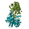
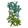
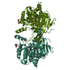
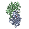
| Title | Crystal structure of Rhizobium etli L-asparaginase ReAV H139A mutant | ||||||
 Components Components | L-asparaginase II protein | ||||||
 Keywords Keywords | HYDROLASE / Rhizobium etli / amidohydrolases / l-asparaginases / zinc-binding proteins / site-directed mutagenesis | ||||||
| Function / homology | L-asparaginase II / L-asparaginase II / metal ion binding / ISOPROPYL ALCOHOL / DI(HYDROXYETHYL)ETHER / L-asparaginase II protein Function and homology information Function and homology information | ||||||
| Biological species |  Rhizobium etli (bacteria) Rhizobium etli (bacteria) | ||||||
| Method |  X-RAY DIFFRACTION / SYNCHROTRON / MOLECULAR REPLACEMENT / Resolution: 1.4 Å X-RAY DIFFRACTION / SYNCHROTRON / MOLECULAR REPLACEMENT / Resolution: 1.4 Å | ||||||
 Authors Authors | Pokrywka, K. / Grzechowiak, M. / Sliwiak, J. / Worsztynowicz, P. / Loch, J.I. / Ruszkowski, M. / Gilski, M. / Jaskolski, M. | ||||||
| Funding support |  Poland, 1items Poland, 1items
| ||||||
 Citation Citation | Journal: Front Chem / Year: 2024 Title: Probing the active site of Class 3 L-asparaginase by mutagenesis. I. Tinkering with the zinc coordination site of ReAV. Authors: Pokrywka, K. / Grzechowiak, M. / Sliwiak, J. / Worsztynowicz, P. / Loch, J.I. / Ruszkowski, M. / Gilski, M. / Jaskolski, M. #1: Journal: IUCrJ / Year: 2021 Title: Structural and biophysical aspects of l-asparaginases: a growing family with amazing diversity. Authors: Loch, J.I. / Jaskolski, M. #2: Journal: Nat Commun / Year: 2021Title: Crystal structures of the elusive Rhizobium etli L-asparaginase reveal a peculiar active site. Authors: Loch, J.I. / Imiolczyk, B. / Sliwiak, J. / Wantuch, A. / Bejger, M. / Gilski, M. / Jaskolski, M. #3: Journal: Acta Crystallogr D Struct Biol / Year: 2023Title: Rhizobium etli has two L-asparaginases with low sequence identity but similar structure and catalytic center. Authors: Loch, J.I. / Worsztynowicz, P. / Sliwiak, J. / Grzechowiak, M. / Imiolczyk, B. / Pokrywka, K. / Chwastyk, M. / Gilski, M. / Jaskolski, M. | ||||||
| History |
|
- Structure visualization
| Structure viewer | Molecule: MolmilJmol/JSmol |
|---|
- Downloads & links
-Download
| PDBx/mmCIF format | 8rue.cif.gz | 575.6 KB | Display | PDBx/mmCIF format |
|---|---|---|---|---|
| PDB format | pdb8rue.ent.gz | 467.1 KB | Display | PDB format |
| PDBx/mmJSON format | 8rue.json.gz | Tree view | PDBx/mmJSON format | |
| Others |  Other downloads Other downloads |
-Validation report
| Arichive directory | https://data.pdbj.org/pub/pdb/validation_reports/ru/8rueftp://data.pdbj.org/pub/pdb/validation_reports/ru/8rue | HTTPS FTP |
|---|
-Related structure data
| Related structure data |  8ruaC  8rudC  8rufC  8rugC C: citing same article ( |
|---|---|
| Similar structure data | |
| Experimental dataset #1 | Data reference: 10.18150/F9YMXN / Data set type: diffraction image data / Metadata reference: 10.18150/F9YMXN |


-Links
 PDBj
PDBj
- Assembly
| Deposited unit | 
| ||||||||||||
|---|---|---|---|---|---|---|---|---|---|---|---|---|---|
| 1 | 
| ||||||||||||
| 2 | 
| ||||||||||||
| Unit cell |
|
-Components
-Protein , 1 types, 4 molecules ABCD
| #1: Protein | Mass: 39487.801 Da / Num. of mol.: 4 / Mutation: H139A Source method: isolated from a genetically manipulated source Details: point mutation H139A / Source: (gene. exp.) Rhizobium etli (bacteria) / Gene: ansA, RHE_PE00350 / Production host: |
|---|
-Non-polymers , 8 types, 1514 molecules 


| #2: Chemical | ChemComp-EDO /  Mass: 62.068 Da / Num. of mol.: 15 / Source method: obtained synthetically / Formula: C2H6O2 Mass: 62.068 Da / Num. of mol.: 15 / Source method: obtained synthetically / Formula: C2H6O2#3: Chemical |  Mass: 92.094 Da / Num. of mol.: 3 / Source method: obtained synthetically / Formula: C3H8O3 Mass: 92.094 Da / Num. of mol.: 3 / Source method: obtained synthetically / Formula: C3H8O3#4: Chemical | ChemComp-IPA /  Mass: 60.095 Da / Num. of mol.: 7 / Source method: obtained synthetically / Formula: C3H8O / Comment: alkaloid*YM Mass: 60.095 Da / Num. of mol.: 7 / Source method: obtained synthetically / Formula: C3H8O / Comment: alkaloid*YM#5: Chemical | ChemComp-ZN /  Mass: 65.409 Da / Num. of mol.: 4 / Source method: obtained synthetically / Formula: Zn / Feature type: SUBJECT OF INVESTIGATION Mass: 65.409 Da / Num. of mol.: 4 / Source method: obtained synthetically / Formula: Zn / Feature type: SUBJECT OF INVESTIGATION#6: Chemical | ChemComp-CL / |  Mass: 35.453 Da / Num. of mol.: 1 / Source method: obtained synthetically / Formula: Cl Mass: 35.453 Da / Num. of mol.: 1 / Source method: obtained synthetically / Formula: Cl#7: Chemical |  Mass: 106.120 Da / Num. of mol.: 3 / Source method: obtained synthetically / Formula: C4H10O3 Mass: 106.120 Da / Num. of mol.: 3 / Source method: obtained synthetically / Formula: C4H10O3#8: Chemical |  Mass: 122.143 Da / Num. of mol.: 2 / Source method: obtained synthetically / Formula: C4H12NO3 / Comment: pH buffer*YM Mass: 122.143 Da / Num. of mol.: 2 / Source method: obtained synthetically / Formula: C4H12NO3 / Comment: pH buffer*YM#9: Water | ChemComp-HOH / | Mass: 18.015 Da / Num. of mol.: 1479 / Source method: isolated from a natural source / Formula: H2O |
|---|
-Details
| Has ligand of interest | Y |
|---|
-Experimental details
-Experiment
| Experiment | Method: X-RAY DIFFRACTION / Number of used crystals: 1 |
|---|
- Sample preparation
| Crystal | Density Matthews: 2.56 Å3/Da / Density % sol: 52.01 % |
|---|---|
| Crystal grow | Temperature: 292 K / Method: vapor diffusion, hanging drop / pH: 9 Details: 22% PEG 3350, 0.2 M MgCl2, 0.1 M Bicine pH 9.0, 1% v/v isopropanol |
-Data collection
| Diffraction | Mean temperature: 100 K / Serial crystal experiment: N |
|---|---|
| Diffraction source | Source: SYNCHROTRON / Site: PETRA III, EMBL c/o DESY  / Beamline: P13 (MX1) / Wavelength: 0.9196 Å / Beamline: P13 (MX1) / Wavelength: 0.9196 Å |
| Detector | Type: DECTRIS PILATUS 6M / Detector: PIXEL / Date: Mar 16, 2023 |
| Radiation | Protocol: SINGLE WAVELENGTH / Monochromatic (M) / Laue (L): M / Scattering type: x-ray |
| Radiation wavelength | Wavelength: 0.9196 Å / Relative weight: 1 |
| Reflection | Resolution: 1.4→77.33 Å / Num. obs: 309393 / % possible obs: 99.2 % / Redundancy: 7 % / Biso Wilson estimate: 15.29 Å2 / CC1/2: 0.999 / Rmerge(I) obs: 0.088 / Rrim(I) all: 0.095 / Net I/σ(I): 12.68 |
| Reflection shell | Resolution: 1.4→1.49 Å / Rmerge(I) obs: 1.322 / Mean I/σ(I) obs: 1.46 / Num. unique obs: 49331 / CC1/2: 0.608 / Rrim(I) all: 1.427 |
- Processing
| Software |
| ||||||||||||||||||||||||||||||||||||||||||||||||||||||||||||||||||||||||||||||||||||
|---|---|---|---|---|---|---|---|---|---|---|---|---|---|---|---|---|---|---|---|---|---|---|---|---|---|---|---|---|---|---|---|---|---|---|---|---|---|---|---|---|---|---|---|---|---|---|---|---|---|---|---|---|---|---|---|---|---|---|---|---|---|---|---|---|---|---|---|---|---|---|---|---|---|---|---|---|---|---|---|---|---|---|---|---|---|
| Refinement | Method to determine structure: MOLECULAR REPLACEMENT / Resolution: 1.4→37.87 Å / SU ML: 0.1514 / Cross valid method: FREE R-VALUE / σ(F): 1.36 / Phase error: 17.7345 Stereochemistry target values: GeoStd + Monomer Library + CDL v1.2 Details: Hydrogen atoms were added at riding position. Anisotropic atomic displacement parameters were used.
| ||||||||||||||||||||||||||||||||||||||||||||||||||||||||||||||||||||||||||||||||||||
| Solvent computation | Shrinkage radii: 0.9 Å / VDW probe radii: 1.1 Å / Solvent model: FLAT BULK SOLVENT MODEL | ||||||||||||||||||||||||||||||||||||||||||||||||||||||||||||||||||||||||||||||||||||
| Displacement parameters | Biso mean: 21.17 Å2 | ||||||||||||||||||||||||||||||||||||||||||||||||||||||||||||||||||||||||||||||||||||
| Refinement step | Cycle: LAST / Resolution: 1.4→37.87 Å
| ||||||||||||||||||||||||||||||||||||||||||||||||||||||||||||||||||||||||||||||||||||
| Refine LS restraints |
| ||||||||||||||||||||||||||||||||||||||||||||||||||||||||||||||||||||||||||||||||||||
| LS refinement shell |
|
