 Movie
Movie Controller

[English] 日本語
 Yorodumi
- PDB-8osw: Crystal structure of Rhizobium etli L-asparaginase ReAIV (R4mC-1) -
+ Open data

- Basic information
| Entry | Database: PDB / ID: 8osw | ||||||
|---|---|---|---|---|---|---|---|
| Title | Crystal structure of Rhizobium etli L-asparaginase ReAIV (R4mC-1) | ||||||
 Components Components | Putative L-asparaginase II protein | ||||||
 Keywords Keywords | HYDROLASE / amidohydrolase / zinc binding protein / structural homology / enzymatic mechanism / enzyme kinetics / occluded water molecules | ||||||
| Function / homology | L-asparaginase II / L-asparaginase II / metal ion binding / L-asparaginase II protein Function and homology information Function and homology information | ||||||
| Biological species |  Rhizobium etli (bacteria) Rhizobium etli (bacteria) | ||||||
| Method |  X-RAY DIFFRACTION / SYNCHROTRON / MOLECULAR REPLACEMENT / Resolution: 1.3 Å X-RAY DIFFRACTION / SYNCHROTRON / MOLECULAR REPLACEMENT / Resolution: 1.3 Å | ||||||
 Authors Authors | Loch, J.I. / Worsztynowicz, P. / Sliwiak, J. / Imiolczyk, B. / Grzechowiak, M. / Gilski, M. / Jaskolski, M. | ||||||
| Funding support |  Poland, 1items Poland, 1items
| ||||||
 Citation Citation | Journal: Acta Crystallogr D Struct Biol / Year: 2023 Title: Rhizobium etli has two L-asparaginases with low sequence identity but similar structure and catalytic center. Authors: Loch, J.I. / Worsztynowicz, P. / Sliwiak, J. / Grzechowiak, M. / Imiolczyk, B. / Pokrywka, K. / Chwastyk, M. / Gilski, M. / Jaskolski, M. | ||||||
| History |
|
- Structure visualization
| Structure viewer | Molecule: MolmilJmol/JSmol |
|---|
- Downloads & links
-Download
| PDBx/mmCIF format | 8osw.cif.gz | 374.4 KB | Display | PDBx/mmCIF format |
|---|---|---|---|---|
| PDB format | pdb8osw.ent.gz | 254.3 KB | Display | PDB format |
| PDBx/mmJSON format | 8osw.json.gz | Tree view | PDBx/mmJSON format | |
| Others |  Other downloads Other downloads |
-Validation report
| Arichive directory | https://data.pdbj.org/pub/pdb/validation_reports/os/8oswftp://data.pdbj.org/pub/pdb/validation_reports/os/8osw | HTTPS FTP |
|---|
-Related structure data
| Related structure data |  8clyC  8clzC  8colC 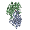 8oriC C: citing same article ( |
|---|---|
| Similar structure data | |
| Experimental dataset #1 | Data reference: 10.18150/NOBOWP / Data set type: diffraction image data / Metadata reference: 10.18150/NOBOWP |
-Links
 PDBj
PDBj- Assembly
| Deposited unit | 
| ||||||||||||
|---|---|---|---|---|---|---|---|---|---|---|---|---|---|
| 1 |
| ||||||||||||
| Unit cell |
|
-Components
| #1: Protein | Mass: 36116.391 Da / Num. of mol.: 2 Source method: isolated from a genetically manipulated source Source: (gene. exp.) Rhizobium etli (bacteria) / Gene: RHE_CH01144 / Production host: #2: Chemical |   Mass: 65.409 Da / Num. of mol.: 2 / Source method: obtained synthetically / Formula: Zn / Feature type: SUBJECT OF INVESTIGATION Mass: 65.409 Da / Num. of mol.: 2 / Source method: obtained synthetically / Formula: Zn / Feature type: SUBJECT OF INVESTIGATION#3: Chemical | ChemComp-CL /   Mass: 35.453 Da / Num. of mol.: 4 / Source method: obtained synthetically / Formula: Cl Mass: 35.453 Da / Num. of mol.: 4 / Source method: obtained synthetically / Formula: Cl#4: Water | ChemComp-HOH / |  Mass: 18.015 Da / Num. of mol.: 871 / Source method: isolated from a natural source / Formula: H2O Mass: 18.015 Da / Num. of mol.: 871 / Source method: isolated from a natural source / Formula: H2OHas ligand of interest | Y | Has protein modification | Y | |
|---|
-Experimental details
-Experiment
| Experiment | Method: X-RAY DIFFRACTION / Number of used crystals: 1 |
|---|
- Sample preparation
| Crystal | Density Matthews: 2.23 Å3/Da / Density % sol: 44.85 % |
|---|---|
| Crystal grow | Temperature: 292 K / Method: vapor diffusion, hanging drop / pH: 7.5 Details: 0.1 M HEPES pH 7.5, 0.2 M MgCl2, 17.5% PEG 8000, 0.3% DDM, 100 mM Asp |
-Data collection
| Diffraction | Mean temperature: 100 K / Serial crystal experiment: N |
|---|---|
| Diffraction source | Source: SYNCHROTRON / Site: PETRA III, EMBL c/o DESY  / Beamline: P13 (MX1) / Wavelength: 0.9762 Å / Beamline: P13 (MX1) / Wavelength: 0.9762 Å |
| Detector | Type: DECTRIS EIGER X 16M / Detector: PIXEL / Date: Jul 8, 2022 |
| Radiation | Protocol: SINGLE WAVELENGTH / Monochromatic (M) / Laue (L): M / Scattering type: x-ray |
| Radiation wavelength | Wavelength: 0.9762 Å / Relative weight: 1 |
| Reflection | Resolution: 1.3→78.35 Å / Num. obs: 150517 / % possible obs: 96.6 % / Redundancy: 6.27 % / Biso Wilson estimate: 13.36 Å2 / CC1/2: 0.998 / Rmerge(I) obs: 0.079 / Rrim(I) all: 0.086 / Net I/σ(I): 14.2 |
| Reflection shell | Resolution: 1.3→1.38 Å / Redundancy: 4.51 % / Rmerge(I) obs: 0.582 / Mean I/σ(I) obs: 2.2 / Num. unique obs: 19954 / CC1/2: 0.791 / Rrim(I) all: 0.655 / % possible all: 79.4 |
- Processing
| Software |
| ||||||||||||||||||||||||||||||||||||||||||||||||||||||||
|---|---|---|---|---|---|---|---|---|---|---|---|---|---|---|---|---|---|---|---|---|---|---|---|---|---|---|---|---|---|---|---|---|---|---|---|---|---|---|---|---|---|---|---|---|---|---|---|---|---|---|---|---|---|---|---|---|---|
| Refinement | Method to determine structure: MOLECULAR REPLACEMENT / Resolution: 1.3→46.2 Å / SU ML: 0.1294 / Cross valid method: FREE R-VALUE / σ(F): 1.35 / Phase error: 17.9913 Stereochemistry target values: GeoStd + Monomer Library + CDL v1.2
| ||||||||||||||||||||||||||||||||||||||||||||||||||||||||
| Solvent computation | Shrinkage radii: 0.9 Å / VDW probe radii: 1.1 Å / Solvent model: FLAT BULK SOLVENT MODEL | ||||||||||||||||||||||||||||||||||||||||||||||||||||||||
| Displacement parameters | Biso mean: 18.37 Å2 | ||||||||||||||||||||||||||||||||||||||||||||||||||||||||
| Refinement step | Cycle: LAST / Resolution: 1.3→46.2 Å
| ||||||||||||||||||||||||||||||||||||||||||||||||||||||||
| Refine LS restraints |
| ||||||||||||||||||||||||||||||||||||||||||||||||||||||||
| LS refinement shell |
|
