 Movie
Movie Controller

[English] 日本語
 Yorodumi
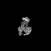
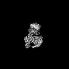
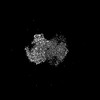
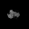
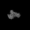
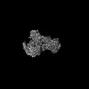
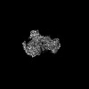
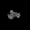
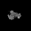
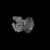
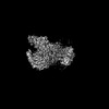
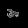
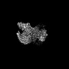
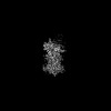
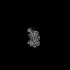
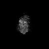
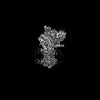
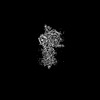
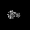
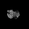
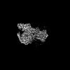
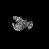
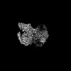
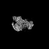
- PDB-8ggp: Locally refined cryoEM structure of receptor from beta-2-adrenerg... -
+ Open data

- Basic information
| Entry | Database: PDB / ID: 8ggp | |||||||||
|---|---|---|---|---|---|---|---|---|---|---|
| Title | Locally refined cryoEM structure of receptor from beta-2-adrenergic receptor in complex with GTP-bound Gs heterotrimer (transition intermediate #8 of 20) | |||||||||
 Components Components | Beta-2 adrenergic receptor | |||||||||
 Keywords Keywords | SIGNALING PROTEIN / GPCR / Adrenergic / Receptor / G protein | |||||||||
| Function / homology |  Function and homology information Function and homology informationbeta2-adrenergic receptor activity / positive regulation of mini excitatory postsynaptic potential / AMPA selective glutamate receptor signaling pathway / heat generation / norepinephrine-epinephrine-mediated vasodilation involved in regulation of systemic arterial blood pressure / adenylate cyclase-inhibiting adrenergic receptor signaling pathway / positive regulation of autophagosome maturation / norepinephrine binding / Adrenoceptors / negative regulation of smooth muscle contraction ...beta2-adrenergic receptor activity / positive regulation of mini excitatory postsynaptic potential / AMPA selective glutamate receptor signaling pathway / heat generation / norepinephrine-epinephrine-mediated vasodilation involved in regulation of systemic arterial blood pressure / adenylate cyclase-inhibiting adrenergic receptor signaling pathway / positive regulation of autophagosome maturation / norepinephrine binding / Adrenoceptors / negative regulation of smooth muscle contraction / positive regulation of cardiac muscle cell contraction / positive regulation of lipophagy / negative regulation of multicellular organism growth / negative regulation of G protein-coupled receptor signaling pathway / smooth muscle contraction / adrenergic receptor signaling pathway / response to psychosocial stress / diet induced thermogenesis / endosome to lysosome transport / positive regulation of cAMP/PKA signal transduction / negative regulation of cardiac muscle cell apoptotic process / adenylate cyclase binding / bone resorption / potassium channel regulator activity / positive regulation of bone mineralization / neuronal dense core vesicle / regulation of sodium ion transport / intercellular bridge / adenylate cyclase-activating adrenergic receptor signaling pathway / positive regulation of cardiac muscle cell apoptotic process / receptor-mediated endocytosis / brown fat cell differentiation / response to cold / clathrin-coated endocytic vesicle membrane / cellular response to amyloid-beta / adenylate cyclase-modulating G protein-coupled receptor signaling pathway / mitotic spindle / Cargo recognition for clathrin-mediated endocytosis / positive regulation of cold-induced thermogenesis / amyloid-beta binding / microtubule cytoskeleton / Clathrin-mediated endocytosis / transcription by RNA polymerase II / G alpha (s) signalling events / positive regulation of MAPK cascade / early endosome / cell surface receptor signaling pathway / lysosome / cilium / signaling receptor complex / endosome / apical plasma membrane / endosome membrane / ciliary basal body / Ub-specific processing proteases / protein-containing complex binding / Golgi apparatus / protein homodimerization activity / positive regulation of transcription by RNA polymerase II / membrane / identical protein binding / nucleus / plasma membrane Similarity search - Function | |||||||||
| Biological species |  Homo sapiens (human) Homo sapiens (human) | |||||||||
| Method | ELECTRON MICROSCOPY / single particle reconstruction / cryo EM / Resolution: 3.2 Å | |||||||||
 Authors Authors | Papasergi-Scott, M.M. / Skiniotis, G. | |||||||||
| Funding support |  United States, 2items United States, 2items
| |||||||||
 Citation Citation | Journal: Nature / Year: 2024 Title: Time-resolved cryo-EM of G-protein activation by a GPCR. Authors: Makaía M Papasergi-Scott / Guillermo Pérez-Hernández / Hossein Batebi / Yang Gao / Gözde Eskici / Alpay B Seven / Ouliana Panova / Daniel Hilger / Marina Casiraghi / Feng He / Luis Maul ...Authors: Makaía M Papasergi-Scott / Guillermo Pérez-Hernández / Hossein Batebi / Yang Gao / Gözde Eskici / Alpay B Seven / Ouliana Panova / Daniel Hilger / Marina Casiraghi / Feng He / Luis Maul / Peter Gmeiner / Brian K Kobilka / Peter W Hildebrand / Georgios Skiniotis /   Abstract: G-protein-coupled receptors (GPCRs) activate heterotrimeric G proteins by stimulating guanine nucleotide exchange in the Gα subunit. To visualize this mechanism, we developed a time-resolved cryo-EM ...G-protein-coupled receptors (GPCRs) activate heterotrimeric G proteins by stimulating guanine nucleotide exchange in the Gα subunit. To visualize this mechanism, we developed a time-resolved cryo-EM approach that examines the progression of ensembles of pre-steady-state intermediates of a GPCR-G-protein complex. By monitoring the transitions of the stimulatory G protein in complex with the β-adrenergic receptor at short sequential time points after GTP addition, we identified the conformational trajectory underlying G-protein activation and functional dissociation from the receptor. Twenty structures generated from sequential overlapping particle subsets along this trajectory, compared to control structures, provide a high-resolution description of the order of main events driving G-protein activation in response to GTP binding. Structural changes propagate from the nucleotide-binding pocket and extend through the GTPase domain, enacting alterations to Gα switch regions and the α5 helix that weaken the G-protein-receptor interface. Molecular dynamics simulations with late structures in the cryo-EM trajectory support that enhanced ordering of GTP on closure of the α-helical domain against the nucleotide-bound Ras-homology domain correlates with α5 helix destabilization and eventual dissociation of the G protein from the GPCR. These findings also highlight the potential of time-resolved cryo-EM as a tool for mechanistic dissection of GPCR signalling events. | |||||||||
| History |
|
- Structure visualization
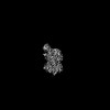
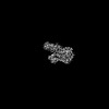
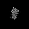
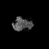
| Structure viewer | Molecule: MolmilJmol/JSmol |
|---|
- Downloads & links
-Download
| PDBx/mmCIF format | 8ggp.cif.gz | 71.6 KB | Display | PDBx/mmCIF format |
|---|---|---|---|---|
| PDB format | pdb8ggp.ent.gz | 47 KB | Display | PDB format |
| PDBx/mmJSON format | 8ggp.json.gz | Tree view | PDBx/mmJSON format | |
| Others |  Other downloads Other downloads |
-Validation report
| Arichive directory | https://data.pdbj.org/pub/pdb/validation_reports/gg/8ggpftp://data.pdbj.org/pub/pdb/validation_reports/gg/8ggp | HTTPS FTP |
|---|
-Related structure data
| Related structure data |  40016MC 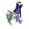 8gdzC 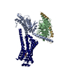 8ge1C 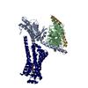 8ge2C 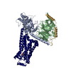 8ge3C 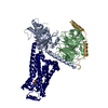 8ge4C 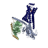 8ge5C 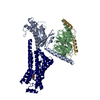 8ge6C 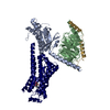 8ge7C 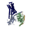 8ge8C 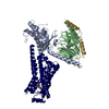 8ge9C 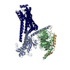 8geaC  8gebC 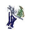 8gecC 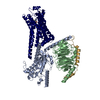 8gedC 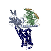 8geeC 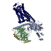 8gefC 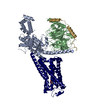 8gegC 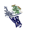 8gehC 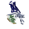 8geiC 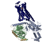 8gejC 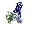 8gfvC 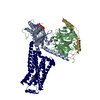 8gfwC 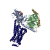 8gfxC 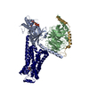 8gfyC 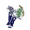 8gfzC 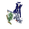 8gg0C  8gg1C 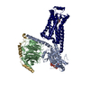 8gg2C 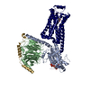 8gg3C  8gg4C 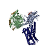 8gg5C 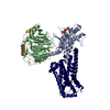 8gg6C 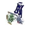 8gg7C 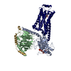 8gg8C 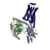 8gg9C 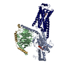 8ggaC 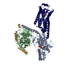 8ggbC  8ggcC 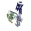 8ggeC 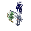 8ggfC 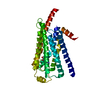 8ggiC 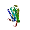 8ggjC 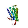 8ggkC 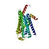 8gglC 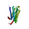 8ggmC 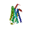 8ggnC 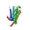 8ggoC 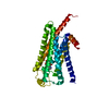 8ggqC  8ggrC  8ggsC  8ggtC  8gguC 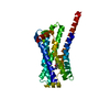 8ggvC 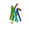 8ggwC 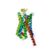 8ggxC 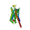 8ggyC 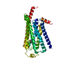 8ggzC 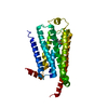 8gh0C 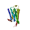 8gh1C  8unlC  8unmC 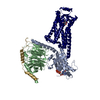 8unnC  8unoC  8unpC  8unqC  8unrC 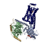 8unsC 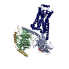 8untC 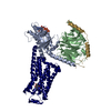 8unuC 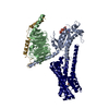 8unvC 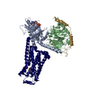 8unwC 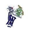 8unxC 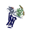 8unyC 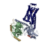 8unzC 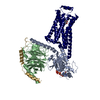 8uo0C 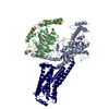 8uo1C 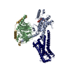 8uo2C  8uo3C  8uo4C C: citing same article ( M: map data used to model this data |
|---|---|
| Similar structure data |


-Links
 PDBj
PDBj


- Assembly
| Deposited unit | 
|
|---|---|
| 1 |
|
-Components
| #1: Protein | Mass: 51767.242 Da / Num. of mol.: 1 Source method: isolated from a genetically manipulated source Source: (gene. exp.) Homo sapiens (human) / Gene: ADRB2, ADRB2R, B2AR / Production host:   Spodoptera frugiperda (fall armyworm) / References: UniProt: P07550 Spodoptera frugiperda (fall armyworm) / References: UniProt: P07550 |
|---|---|
| #2: Chemical | ChemComp-G1I / (  Mass: 209.242 Da / Num. of mol.: 1 / Source method: obtained synthetically / Formula: C11H15NO3 Mass: 209.242 Da / Num. of mol.: 1 / Source method: obtained synthetically / Formula: C11H15NO3 |
| Has ligand of interest | N |
| Has protein modification | Y |
-Experimental details
-Experiment
| Experiment | Method: ELECTRON MICROSCOPY |
|---|---|
| EM experiment | Aggregation state: PARTICLE / 3D reconstruction method: single particle reconstruction |
- Sample preparation
| Component | Name: Complex of beta-2 adrenergic receptor and Gs heterotrimer with GTP Type: COMPLEX Details: Combined datasets of the protein complex from samples plunge frozen at 5 sec, 10 sec, or 17 sec after GTP addition to the sample. Entity ID: #1 / Source: RECOMBINANT |
|---|---|
| Molecular weight | Experimental value: NO |
| Source (natural) | Organism: Homo sapiens (human) |
| Source (recombinant) | Organism: Spodoptera frugiperda (fall armyworm) |
| Buffer solution | pH: 7.5 Details: GTP was added just prior to freezing at 5 sec, 10 sec, or 17 sec before plunging. |
| Specimen | Embedding applied: NO / Shadowing applied: NO / Staining applied: NO / Vitrification applied: YES |
| Specimen support | Grid type: UltrAuFoil R1.2/1.3 |
| Vitrification | Instrument: FEI VITROBOT MARK IV / Cryogen name: ETHANE / Humidity: 100 % / Chamber temperature: 277.15 K Details: GTP was added just prior to freezing at 5 sec, 10 sec, or 17 sec before plunging. |
- Electron microscopy imaging
| Experimental equipment |  Model: Titan Krios / Image courtesy: FEI Company |
|---|---|
| Microscopy | Model: FEI TITAN KRIOS |
| Electron gun | Electron source:  FIELD EMISSION GUN / Accelerating voltage: 300 kV / Illumination mode: FLOOD BEAM FIELD EMISSION GUN / Accelerating voltage: 300 kV / Illumination mode: FLOOD BEAM |
| Electron lens | Mode: BRIGHT FIELD / Nominal defocus max: 2000 nm / Nominal defocus min: 400 nm |
| Specimen holder | Cryogen: NITROGEN / Specimen holder model: FEI TITAN KRIOS AUTOGRID HOLDER |
| Image recording | Electron dose: 50 e/Å2 / Detector mode: COUNTING / Film or detector model: GATAN K3 (6k x 4k) Details: The map is the result of combining multiple datasets: cryo-EM imaging of the beta-2AR-Gs + GTP (5 sec) complex was performed on a Titan Krios electron microscope equipped with a K3 Summit ...Details: The map is the result of combining multiple datasets: cryo-EM imaging of the beta-2AR-Gs + GTP (5 sec) complex was performed on a Titan Krios electron microscope equipped with a K3 Summit direct electron detector (Gatan). The microscope was operated at 300 kV accelerating voltage, with a nominal magnification of 105,000x in counting mode resulting in a magnified pixel size of 0.8677 Angstrom. A total exposure of 60.48 electrons/ Angstrom^2 over 63 frames with defocus ranging from -1.0 - -2.0 micrometers was used. Cryo-EM imaging of beta-2AR-Gs + GTP (10 sec) complex was performed on four separate grids over three collection sessions. The microscope was operated at 300 kV accelerating voltage, with a magnification at the camera of 58,679x in counting mode resulting in a magnified pixel size of 0.8521 Angstrom. For the first and second grids, movies were obtained at an exposure rate of 21.13 electrons/Angstrum^2/sec with defocus ranging from -0.4 - -2.0 micrometers. The total exposure time was 2.717 sec over 77 frames per movie stack. For an additional collection of the first grid, movies were obtained at an exposure rate of 20.95 electrons/ Angstrum^2/sec with defocus ranging from -0.4 -2.0 micrometers. The total exposure time was 2.717 sec over 77 frames per movie stack. For the third and fourth grids, movies were obtained at an exposure rate of 30.71 electrons/ Angstrum^2/sec with defocus ranging from -0.5 - -1.6 micrometers. The total exposure time was 2.008 sec over 79 frames per movie stack. Cryo-EM imaging of beta-2AR-Gs + GTP (17 sec) was performed on a Titan Krios equipped with a post-column energy filter, with a magnification of 105,000x in counting mode resulting in a magnified pixel size of 0.8677 Angstrom. Movies were obtained at an exposure rate of 32.46 electrons/Angstrum^2/sec with defocus ranging from -0.4 - -0.9 micrometers. The total exposure time was 1.999 sec over 79 frames per movie stack. |
- Processing
| EM software |
| ||||||||||||||||||||||||||||
|---|---|---|---|---|---|---|---|---|---|---|---|---|---|---|---|---|---|---|---|---|---|---|---|---|---|---|---|---|---|
| CTF correction | Type: NONE | ||||||||||||||||||||||||||||
| Particle selection | Num. of particles selected: 19918625 | ||||||||||||||||||||||||||||
| 3D reconstruction | Resolution: 3.2 Å / Resolution method: FSC 0.143 CUT-OFF / Num. of particles: 241817 / Symmetry type: POINT | ||||||||||||||||||||||||||||
| Refine LS restraints |
|
