 Movie
Movie Controller

[English] 日本語
 Yorodumi
- PDB-8eia: Crystal structure of beta-catenin and the MDM2 p53-binding domain... -
+ Open data

- Basic information
| Entry | Database: PDB / ID: 8eia | ||||||
|---|---|---|---|---|---|---|---|
| Title | Crystal structure of beta-catenin and the MDM2 p53-binding domain in complex with H333, a Helicon Polypeptide | ||||||
 Components Components |
| ||||||
 Keywords Keywords | LIGASE / E3 ligase / complex / stapled peptide | ||||||
| Function / homology |  Function and homology information Function and homology informationpositive regulation of heparan sulfate proteoglycan biosynthetic process / cranial ganglion development / beta-catenin-ICAT complex / CDH11 homotypic and heterotypic interactions / embryonic skeletal limb joint morphogenesis / Regulation of CDH19 Expression and Function / astrocyte-dopaminergic neuron signaling / beta-catenin-TCF7L2 complex / regulation of nephron tubule epithelial cell differentiation / regulation of timing of anagen ...positive regulation of heparan sulfate proteoglycan biosynthetic process / cranial ganglion development / beta-catenin-ICAT complex / CDH11 homotypic and heterotypic interactions / embryonic skeletal limb joint morphogenesis / Regulation of CDH19 Expression and Function / astrocyte-dopaminergic neuron signaling / beta-catenin-TCF7L2 complex / regulation of nephron tubule epithelial cell differentiation / regulation of timing of anagen / negative regulation of mitotic cell cycle, embryonic / Binding of TCF/LEF:CTNNB1 to target gene promoters / regulation of centriole-centriole cohesion / RUNX3 regulates WNT signaling / regulation of centromeric sister chromatid cohesion / Regulation of CDH11 function / regulation of fibroblast proliferation / Scrib-APC-beta-catenin complex / beta-catenin-TCF complex / Specification of the neural plate border / positive regulation of skeletal muscle tissue development / synaptic vesicle clustering / Formation of the nephric duct / endothelial tube morphogenesis / hindbrain development / dorsal root ganglion development / fascia adherens / mesenchymal to epithelial transition / cranial skeletal system development / sympathetic ganglion development / cellular response to vitamin B1 / response to formaldehyde / presynaptic active zone cytoplasmic component / regulation of protein localization to cell surface / response to ether / response to water-immersion restraint stress / mesenchymal stem cell differentiation / detection of muscle stretch / positive regulation of odontoblast differentiation / cellular response to UV-C / regulation of epithelial to mesenchymal transition / alpha-catenin binding / cellular response to indole-3-methanol / histone methyltransferase binding / regulation of calcium ion import / apicolateral plasma membrane / positive regulation of homotypic cell-cell adhesion / neuron projection extension / Germ layer formation at gastrulation / hair cell differentiation / fibroblast activation / cell-cell adhesion mediated by cadherin / regulation of protein catabolic process at postsynapse, modulating synaptic transmission / flotillin complex / Formation of definitive endoderm / Trafficking of AMPA receptors / regulation of smooth muscle cell proliferation / Formation of axial mesoderm / beta-catenin destruction complex / negative regulation of protein sumoylation / embryonic brain development / Apoptotic cleavage of cell adhesion proteins / catenin complex / midbrain dopaminergic neuron differentiation / LRR FLII-interacting protein 1 (LRRFIP1) activates type I IFN production / positive regulation of blood vessel branching / protein localization to cell surface / receptor serine/threonine kinase binding / Beta-catenin phosphorylation cascade / Signaling by GSK3beta mutants / CTNNB1 S33 mutants aren't phosphorylated / CTNNB1 S37 mutants aren't phosphorylated / CTNNB1 S45 mutants aren't phosphorylated / CTNNB1 T41 mutants aren't phosphorylated / negative regulation of oxidative stress-induced neuron intrinsic apoptotic signaling pathway / negative regulation of intrinsic apoptotic signaling pathway by p53 class mediator / cellular response to alkaloid / I-SMAD binding / Regulation of CDH1 Function / SUMO transferase activity / response to steroid hormone / negative regulation of protein processing / positive regulation of vascular associated smooth muscle cell migration / Adherens junctions interactions / Wnt signalosome / long-term synaptic depression / peroxisome proliferator activated receptor binding / AKT phosphorylates targets in the cytosol / positive regulation of neuroblast proliferation / response to iron ion / Cardiogenesis / adherens junction assembly / Disassembly of the destruction complex and recruitment of AXIN to the membrane / NEDD8 ligase activity / cellular response to peptide hormone stimulus / regulation of protein catabolic process / positive regulation of muscle cell differentiation / stem cell population maintenance / ligase activity / cellular response to antibiotic Similarity search - Function | ||||||
| Biological species |  Homo sapiens (human) Homo sapiens (human)synthetic construct (others) | ||||||
| Method |  X-RAY DIFFRACTION / SYNCHROTRON / MOLECULAR REPLACEMENT / Resolution: 3.6 Å X-RAY DIFFRACTION / SYNCHROTRON / MOLECULAR REPLACEMENT / Resolution: 3.6 Å | ||||||
 Authors Authors | Li, K. / Travaline, T.L. / Swiecicki, J.-M. / Tokareva, O.S. / Thomson, T.M. / Verdine, G.L. / McGee, J.H. | ||||||
| Funding support |  United States, 1items United States, 1items
| ||||||
 Citation Citation | Journal: Nat Commun / Year: 2023 Title: Recognition and reprogramming of E3 ubiquitin ligase surfaces by alpha-helical peptides. Authors: Tokareva, O.S. / Li, K. / Travaline, T.L. / Thomson, T.M. / Swiecicki, J.M. / Moussa, M. / Ramirez, J.D. / Litchman, S. / Verdine, G.L. / McGee, J.H. | ||||||
| History |
|
- Structure visualization
| Structure viewer | Molecule: MolmilJmol/JSmol |
|---|
- Downloads & links
-Download
| PDBx/mmCIF format | 8eia.cif.gz | 282.4 KB | Display | PDBx/mmCIF format |
|---|---|---|---|---|
| PDB format | pdb8eia.ent.gz | 203.6 KB | Display | PDB format |
| PDBx/mmJSON format | 8eia.json.gz | Tree view | PDBx/mmJSON format | |
| Others |  Other downloads Other downloads |
-Validation report
| Arichive directory | https://data.pdbj.org/pub/pdb/validation_reports/ei/8eiaftp://data.pdbj.org/pub/pdb/validation_reports/ei/8eia | HTTPS FTP |
|---|
-Related structure data
| Related structure data |  8ehzC  8ei0C  8ei1C  8ei2C  8ei3C  8ei4C  8ei5C  8ei6C  8ei7C  8ei8C  8ei9C  8eibC 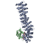 8eicC 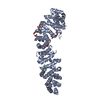 7uwiS S: Starting model for refinement C: citing same article ( |
|---|---|
| Similar structure data |
-Links
 PDBj
PDBj

















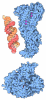
- Assembly
| Deposited unit | 
| ||||||||||||
|---|---|---|---|---|---|---|---|---|---|---|---|---|---|
| 1 |
| ||||||||||||
| Unit cell |
|
-Components
| #1: Protein | Mass: 58139.312 Da / Num. of mol.: 1 Source method: isolated from a genetically manipulated source Source: (gene. exp.) Homo sapiens (human) / Gene: CTNNB1, CTNNB, OK/SW-cl.35, PRO2286 / Production host:   Escherichia coli (E. coli) / References: UniProt: P35222 Escherichia coli (E. coli) / References: UniProt: P35222 |
|---|---|
| #2: Protein | Mass: 11099.000 Da / Num. of mol.: 1 / Fragment: P53 binding domain Source method: isolated from a genetically manipulated source Source: (gene. exp.) Homo sapiens (human) / Gene: MDM2 / Production host: Escherichia coli (E. coli)References: UniProt: Q00987, RING-type E3 ubiquitin transferase |
| #3: Protein/peptide | Mass: 2623.918 Da / Num. of mol.: 1 / Source method: obtained synthetically / Source: (synth.) synthetic construct (others) |
| #4: Chemical | ChemComp-WHL /   Mass: 192.214 Da / Num. of mol.: 1 / Source method: obtained synthetically / Formula: C10H12N2O2 Mass: 192.214 Da / Num. of mol.: 1 / Source method: obtained synthetically / Formula: C10H12N2O2 |
| Has ligand of interest | N |
| Has protein modification | Y |
-Experimental details
-Experiment
| Experiment | Method: X-RAY DIFFRACTION / Number of used crystals: 1 |
|---|
- Sample preparation
| Crystal | Density Matthews: 2.45 Å3/Da / Density % sol: 49.79 % |
|---|---|
| Crystal grow | Temperature: 291 K / Method: vapor diffusion, sitting drop / pH: 7 Details: 0.1 M Potassium chloride, 0.1 M HEPES pH 7, 15% w/v PEG 5000 MME |
-Data collection
| Diffraction | Mean temperature: 100 K / Serial crystal experiment: N |
|---|---|
| Diffraction source | Source: SYNCHROTRON / Site: NSLS-II / Beamline: 17-ID-1 / Wavelength: 0.9201 Å |
| Detector | Type: DECTRIS EIGER X 9M / Detector: PIXEL / Date: Dec 11, 2021 |
| Radiation | Protocol: SINGLE WAVELENGTH / Monochromatic (M) / Laue (L): M / Scattering type: x-ray |
| Radiation wavelength | Wavelength: 0.9201 Å / Relative weight: 1 |
| Reflection | Resolution: 3.6→48.03 Å / Num. obs: 7517 / % possible obs: 92.31 % / Redundancy: 5.5 % / Biso Wilson estimate: 86.83 Å2 / CC1/2: 0.978 / Rmerge(I) obs: 0.241 / Net I/σ(I): 5.3 |
| Reflection shell | Resolution: 3.6→3.94 Å / Redundancy: 5.4 % / Rmerge(I) obs: 0.912 / Mean I/σ(I) obs: 1.7 / Num. unique obs: 1803 / CC1/2: 0.491 / % possible all: 94.29 |
- Processing
| Software |
| |||||||||||||||||||||||||||||||||||||||||||||||||||||||||||||||||||||||||||||
|---|---|---|---|---|---|---|---|---|---|---|---|---|---|---|---|---|---|---|---|---|---|---|---|---|---|---|---|---|---|---|---|---|---|---|---|---|---|---|---|---|---|---|---|---|---|---|---|---|---|---|---|---|---|---|---|---|---|---|---|---|---|---|---|---|---|---|---|---|---|---|---|---|---|---|---|---|---|---|
| Refinement | Method to determine structure: MOLECULAR REPLACEMENT Starting model: 7UWI Resolution: 3.6→47.73 Å / SU ML: 0.6739 / Cross valid method: FREE R-VALUE / σ(F): 1.35 / Phase error: 41.6938 Stereochemistry target values: GeoStd + Monomer Library + CDL v1.2
| |||||||||||||||||||||||||||||||||||||||||||||||||||||||||||||||||||||||||||||
| Solvent computation | Shrinkage radii: 0.9 Å / VDW probe radii: 1.1 Å / Solvent model: FLAT BULK SOLVENT MODEL | |||||||||||||||||||||||||||||||||||||||||||||||||||||||||||||||||||||||||||||
| Displacement parameters | Biso mean: 100.55 Å2 | |||||||||||||||||||||||||||||||||||||||||||||||||||||||||||||||||||||||||||||
| Refinement step | Cycle: LAST / Resolution: 3.6→47.73 Å
| |||||||||||||||||||||||||||||||||||||||||||||||||||||||||||||||||||||||||||||
| Refine LS restraints |
| |||||||||||||||||||||||||||||||||||||||||||||||||||||||||||||||||||||||||||||
| LS refinement shell |
| |||||||||||||||||||||||||||||||||||||||||||||||||||||||||||||||||||||||||||||
| Refinement TLS params. | Method: refined / Origin x: 7.74981909216 Å / Origin y: 13.7179774078 Å / Origin z: 20.7883535427 Å
| |||||||||||||||||||||||||||||||||||||||||||||||||||||||||||||||||||||||||||||
| Refinement TLS group | Selection details: all |
