 Movie
Movie Controller

[English] 日本語
 Yorodumi
- PDB-8ei2: Crystal structure of the N-terminal domain of CUL5 in complex wit... -
+ Open data

- Basic information
| Entry | Database: PDB / ID: 8ei2 | ||||||
|---|---|---|---|---|---|---|---|
| Title | Crystal structure of the N-terminal domain of CUL5 in complex with H314, a Helicon Polypeptide | ||||||
 Components Components |
| ||||||
 Keywords Keywords | LIGASE / E3 ligase / complex / stapled peptide | ||||||
| Function / homology |  Function and homology information Function and homology informationnegative regulation of focal adhesion disassembly / ERBB2 signaling pathway / negative regulation of focal adhesion assembly / reelin-mediated signaling pathway / regulation of neuron migration / protein K11-linked ubiquitination / Cul5-RING ubiquitin ligase complex / SCF ubiquitin ligase complex / SCF-dependent proteasomal ubiquitin-dependent protein catabolic process / ubiquitin ligase complex scaffold activity ...negative regulation of focal adhesion disassembly / ERBB2 signaling pathway / negative regulation of focal adhesion assembly / reelin-mediated signaling pathway / regulation of neuron migration / protein K11-linked ubiquitination / Cul5-RING ubiquitin ligase complex / SCF ubiquitin ligase complex / SCF-dependent proteasomal ubiquitin-dependent protein catabolic process / ubiquitin ligase complex scaffold activity / site of DNA damage / intrinsic apoptotic signaling pathway / G1/S transition of mitotic cell cycle / Vif-mediated degradation of APOBEC3G / Inactivation of CSF3 (G-CSF) signaling / Evasion by RSV of host interferon responses / calcium channel activity / Downregulation of ERBB2 signaling / ubiquitin-protein transferase activity / cell migration / Antigen processing: Ubiquitination & Proteasome degradation / signaling receptor activity / Neddylation / proteasome-mediated ubiquitin-dependent protein catabolic process / protein-macromolecule adaptor activity / positive regulation of cell migration / protein ubiquitination / ubiquitin protein ligase binding / nucleus / cytoplasm / cytosol Similarity search - Function | ||||||
| Biological species |  Homo sapiens (human) Homo sapiens (human)synthetic construct (others) | ||||||
| Method |  X-RAY DIFFRACTION / SYNCHROTRON / MOLECULAR REPLACEMENT / Resolution: 2.8 Å X-RAY DIFFRACTION / SYNCHROTRON / MOLECULAR REPLACEMENT / Resolution: 2.8 Å | ||||||
 Authors Authors | Li, K. / Tokareva, O.S. / Thomson, T.M. / Verdine, G.L. / McGee, J.H. | ||||||
| Funding support |  United States, 1items United States, 1items
| ||||||
 Citation Citation | Journal: Nat Commun / Year: 2023 Title: Recognition and reprogramming of E3 ubiquitin ligase surfaces by alpha-helical peptides. Authors: Tokareva, O.S. / Li, K. / Travaline, T.L. / Thomson, T.M. / Swiecicki, J.M. / Moussa, M. / Ramirez, J.D. / Litchman, S. / Verdine, G.L. / McGee, J.H. | ||||||
| History |
|
- Structure visualization
| Structure viewer | Molecule: MolmilJmol/JSmol |
|---|
- Downloads & links
-Download
| PDBx/mmCIF format | 8ei2.cif.gz | 96.6 KB | Display | PDBx/mmCIF format |
|---|---|---|---|---|
| PDB format | pdb8ei2.ent.gz | 66.9 KB | Display | PDB format |
| PDBx/mmJSON format | 8ei2.json.gz | Tree view | PDBx/mmJSON format | |
| Others |  Other downloads Other downloads |
-Validation report
| Arichive directory | https://data.pdbj.org/pub/pdb/validation_reports/ei/8ei2ftp://data.pdbj.org/pub/pdb/validation_reports/ei/8ei2 | HTTPS FTP |
|---|
-Related structure data
| Related structure data | 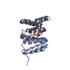 8ehzC  8ei0C 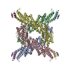 8ei1C  8ei3C  8ei4C 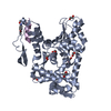 8ei5C 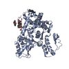 8ei6C 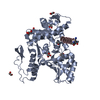 8ei7C 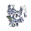 8ei8C 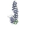 8ei9C 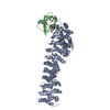 8eiaC  8eibC 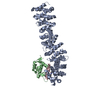 8eicC  2wzkS S: Starting model for refinement C: citing same article ( |
|---|---|
| Similar structure data |
-Links
 PDBj
PDBj
- Assembly
| Deposited unit | 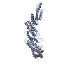
| ||||||||||
|---|---|---|---|---|---|---|---|---|---|---|---|
| 1 |
| ||||||||||
| Unit cell |
|
-Components
| #1: Protein | Mass: 44972.461 Da / Num. of mol.: 1 / Fragment: N-terminal domain Source method: isolated from a genetically manipulated source Source: (gene. exp.) Homo sapiens (human) / Gene: CUL5, VACM1 / Production host:  |
|---|---|
| #2: Protein/peptide | Mass: 1872.065 Da / Num. of mol.: 1 / Source method: obtained synthetically / Source: (synth.) synthetic construct (others) |
| #3: Chemical | ChemComp-WHL /   Mass: 192.214 Da / Num. of mol.: 1 / Source method: obtained synthetically / Formula: C10H12N2O2 Mass: 192.214 Da / Num. of mol.: 1 / Source method: obtained synthetically / Formula: C10H12N2O2 |
| Has ligand of interest | N |
| Has protein modification | Y |
-Experimental details
-Experiment
| Experiment | Method: X-RAY DIFFRACTION / Number of used crystals: 1 |
|---|
- Sample preparation
| Crystal | Density Matthews: 5.3 Å3/Da / Density % sol: 76.8 % |
|---|---|
| Crystal grow | Temperature: 291 K / Method: vapor diffusion, sitting drop / pH: 6.5 Details: 30% w/v PEG 8000, 0.1M MES Sodium Salt pH6.5, 0.2M Ammonium Sulfate, 4% v/v 1,3-Propanediol |
-Data collection
| Diffraction | Mean temperature: 100 K / Serial crystal experiment: N |
|---|---|
| Diffraction source | Source: SYNCHROTRON / Site: SPring-8  / Beamline: BL45XU / Wavelength: 1 Å / Beamline: BL45XU / Wavelength: 1 Å |
| Detector | Type: DECTRIS PILATUS 6M / Detector: PIXEL / Date: Nov 12, 2020 |
| Radiation | Protocol: SINGLE WAVELENGTH / Monochromatic (M) / Laue (L): M / Scattering type: x-ray |
| Radiation wavelength | Wavelength: 1 Å / Relative weight: 1 |
| Reflection twin | Operator: h,-h-k,-l / Fraction: 0.85 |
| Reflection | Resolution: 2.8→47.88 Å / Num. obs: 24154 / % possible obs: 98.2 % / Redundancy: 13.2 % / Biso Wilson estimate: -4.64 Å2 / CC1/2: 0.988 / Rmerge(I) obs: 0.322 / Net I/σ(I): 5 |
| Reflection shell | Resolution: 2.8→2.95 Å / Redundancy: 13.7 % / Mean I/σ(I) obs: 1.8 / Num. unique obs: 3518 / CC1/2: 0.888 / % possible all: 99.3 |
- Processing
| Software |
| |||||||||||||||||||||||||||||||||||||||||||||||||
|---|---|---|---|---|---|---|---|---|---|---|---|---|---|---|---|---|---|---|---|---|---|---|---|---|---|---|---|---|---|---|---|---|---|---|---|---|---|---|---|---|---|---|---|---|---|---|---|---|---|---|
| Refinement | Method to determine structure: MOLECULAR REPLACEMENT Starting model: 2WZK Resolution: 2.8→47.88 Å / SU ML: 0.4852 / Cross valid method: FREE R-VALUE / σ(F): 1.36 / Phase error: 33.6178 Stereochemistry target values: GeoStd + Monomer Library + CDL v1.2
| |||||||||||||||||||||||||||||||||||||||||||||||||
| Solvent computation | Shrinkage radii: 0.9 Å / VDW probe radii: 1.11 Å / Solvent model: FLAT BULK SOLVENT MODEL | |||||||||||||||||||||||||||||||||||||||||||||||||
| Displacement parameters | Biso mean: 20.32 Å2 | |||||||||||||||||||||||||||||||||||||||||||||||||
| Refinement step | Cycle: LAST / Resolution: 2.8→47.88 Å
| |||||||||||||||||||||||||||||||||||||||||||||||||
| Refine LS restraints |
| |||||||||||||||||||||||||||||||||||||||||||||||||
| LS refinement shell |
|
