Movie
Movie Controller

[English] 日本語
 Yorodumi
- PDB-8axk: Type 3 secretion system export apparatus core, inner rod and need... -
+ Open data

- Basic information
| Entry | Database: PDB / ID: 8axk | ||||||
|---|---|---|---|---|---|---|---|
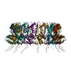
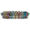
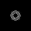
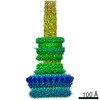
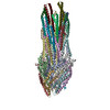
| Title | Type 3 secretion system export apparatus core, inner rod and needle of Shigella flexneri | ||||||
 Components Components |
| ||||||
 Keywords Keywords | PROTEIN TRANSPORT / type 3 secretion / needle complex / virulence factor / shigella / infection | ||||||
| Function / homology |  Function and homology information Function and homology informationtype III protein secretion system complex / type II protein secretion system complex / protein secretion by the type III secretion system / protein secretion / protein targeting / cell outer membrane / protein transport / cell surface / extracellular region / identical protein binding / plasma membrane Similarity search - Function | ||||||
| Biological species |  Shigella flexneri (bacteria) Shigella flexneri (bacteria) | ||||||
| Method | ELECTRON MICROSCOPY / single particle reconstruction / cryo EM / Resolution: 4.05 Å | ||||||
 Authors Authors | Lunelli, M. | ||||||
| Funding support |  Germany, 1items Germany, 1items
| ||||||
 Citation Citation | Journal: Protein Sci / Year: 2023 Title: Integrative structural analysis of the type III secretion system needle complex from Shigella flexneri. Authors: Lara Flacht / Michele Lunelli / Karol Kaszuba / Zhuo Angel Chen / Francis J O' Reilly / Juri Rappsilber / Jan Kosinski / Michael Kolbe /  Abstract: The type III secretion system (T3SS) is a large, transmembrane protein machinery used by various pathogenic gram-negative bacteria to transport virulence factors into the host cell during infection. ...The type III secretion system (T3SS) is a large, transmembrane protein machinery used by various pathogenic gram-negative bacteria to transport virulence factors into the host cell during infection. Understanding the structure of T3SSs is crucial for future developments of therapeutics that could target this system. However, much of the knowledge about the structure of T3SS is available only for Salmonella, and it is unclear how this large assembly is conserved across species. Here, we combined cryo-electron microscopy, cross-linking mass spectrometry, and integrative modeling to determine the structure of the T3SS needle complex from Shigella flexneri. We show that the Shigella T3SS exhibits unique features distinguishing it from other structurally characterized T3SSs. The secretin pore complex adopts a new fold of its C-terminal S domain and the pilotin MxiM[SctG] locates around the outer surface of the pore. The export apparatus structure exhibits a conserved pseudohelical arrangement but includes the N-terminal domain of the SpaS[SctU] subunit, which was not present in any of the previously published virulence-related T3SS structures. Similar to other T3SSs, however, the apparatus is anchored within the needle complex by a network of flexible linkers that either adjust conformation to connect to equivalent patches on the secretin oligomer or bind distinct surface patches at the same height of the export apparatus. The conserved and unique features delineated by our analysis highlight the necessity to analyze T3SS in a species-specific manner, in order to fully understand the underlying molecular mechanisms of these systems. The structure of the type III secretion system from Shigella flexneri delineates conserved and unique features, which could be used for the development of broad-range therapeutics. | ||||||
| History |
|
- Structure visualization
| Structure viewer | Molecule: MolmilJmol/JSmol |
|---|
- Downloads & links
-Download
| PDBx/mmCIF format | 8axk.cif.gz | 1.1 MB | Display | PDBx/mmCIF format |
|---|---|---|---|---|
| PDB format | pdb8axk.ent.gz | Display | PDB format | |
| PDBx/mmJSON format | 8axk.json.gz | Tree view | PDBx/mmJSON format | |
| Others |  Other downloads Other downloads |
-Validation report
| Arichive directory | https://data.pdbj.org/pub/pdb/validation_reports/ax/8axkftp://data.pdbj.org/pub/pdb/validation_reports/ax/8axk | HTTPS FTP |
|---|
-Related structure data
| Related structure data |  15700MC  8axlC  8axnC M: map data used to model this data C: citing same article ( |
|---|---|
| Similar structure data |


-Links
 PDBj
PDBj


- Assembly
| Deposited unit | 
|
|---|---|
| 1 |
|
-Components
-Surface presentation of antigens protein ... , 4 types, 11 molecules ABCDEFGHIJK
| #1: Protein | Mass: 24215.562 Da / Num. of mol.: 5 / Source method: isolated from a natural source / Source: (natural) Shigella flexneri (bacteria) / References: UniProt: P0A1L3#2: Protein | | Mass: 28513.773 Da / Num. of mol.: 1 / Source method: isolated from a natural source / Source: (natural) Shigella flexneri (bacteria) / References: UniProt: P0A1M6#3: Protein | Mass: 9433.338 Da / Num. of mol.: 4 / Source method: isolated from a natural source / Source: (natural) Shigella flexneri (bacteria) / References: UniProt: P0A1M4#4: Protein | | Mass: 39903.348 Da / Num. of mol.: 1 / Source method: isolated from a natural source / Source: (natural) Shigella flexneri (bacteria) / References: UniProt: P0A1M8 |
|---|
-Protein , 4 types, 74 molecules MNOPQRSTUVWabcdefghijklmnopqrs...
| #5: Protein | Mass: 10640.000 Da / Num. of mol.: 6 / Source method: isolated from a natural source / Source: (natural) Shigella flexneri (bacteria) / References: UniProt: P0A225#6: Protein | Mass: 11060.279 Da / Num. of mol.: 28 Source method: isolated from a genetically manipulated source Source: (gene. exp.) Shigella flexneri (bacteria) / Gene: mxiH, CP0137 / Plasmid: pASK-IBA5plus / Production host: Shigella flexneri (bacteria) / References: UniProt: P0A223#7: Protein | Mass: 63230.414 Da / Num. of mol.: 16 / Source method: isolated from a natural source / Source: (natural) Shigella flexneri (bacteria) / References: UniProt: Q04641#8: Protein | Mass: 27542.055 Da / Num. of mol.: 24 / Source method: isolated from a natural source / Source: (natural) Shigella flexneri (bacteria) / References: UniProt: Q06081 |
|---|
-Details
| Has protein modification | Y |
|---|
-Experimental details
-Experiment
| Experiment | Method: ELECTRON MICROSCOPY |
|---|---|
| EM experiment | Aggregation state: PARTICLE / 3D reconstruction method: single particle reconstruction |
- Sample preparation
| Component |
| ||||||||||||||||||||||||||||||||||||||||||
|---|---|---|---|---|---|---|---|---|---|---|---|---|---|---|---|---|---|---|---|---|---|---|---|---|---|---|---|---|---|---|---|---|---|---|---|---|---|---|---|---|---|---|---|
| Molecular weight |
| ||||||||||||||||||||||||||||||||||||||||||
| Source (natural) |
| ||||||||||||||||||||||||||||||||||||||||||
| Buffer solution | pH: 8 | ||||||||||||||||||||||||||||||||||||||||||
| Specimen | Embedding applied: NO / Shadowing applied: NO / Staining applied: NO / Vitrification applied: YES | ||||||||||||||||||||||||||||||||||||||||||
| Specimen support | Grid material: COPPER / Grid type: Quantifoil R2/1 | ||||||||||||||||||||||||||||||||||||||||||
| Vitrification | Instrument: FEI VITROBOT MARK IV / Cryogen name: ETHANE / Humidity: 100 % / Chamber temperature: 295 K |
- Electron microscopy imaging
| Experimental equipment |  Model: Titan Krios / Image courtesy: FEI Company |
|---|---|
| Microscopy | Model: FEI TITAN KRIOS |
| Electron gun | Electron source:  FIELD EMISSION GUN / Accelerating voltage: 300 kV / Illumination mode: OTHER FIELD EMISSION GUN / Accelerating voltage: 300 kV / Illumination mode: OTHER |
| Electron lens | Mode: BRIGHT FIELD / Nominal magnification: 101179 X / Nominal defocus max: 4000 nm / Nominal defocus min: 1500 nm / Cs: 2.7 mm |
| Specimen holder | Cryogen: NITROGEN / Specimen holder model: FEI TITAN KRIOS AUTOGRID HOLDER |
| Image recording | Average exposure time: 1.5 sec. / Electron dose: 25 e/Å2 / Detector mode: INTEGRATING / Film or detector model: FEI FALCON II (4k x 4k) / Num. of grids imaged: 1 / Num. of real images: 5238 |
| Image scans | Width: 4096 / Height: 4096 / Movie frames/image: 7 / Used frames/image: 1-6 |
- Processing
| Software | Name: PHENIX / Version: 1.18.2_3874: / Classification: refinement | ||||||||||||||||||||||||||||||||
|---|---|---|---|---|---|---|---|---|---|---|---|---|---|---|---|---|---|---|---|---|---|---|---|---|---|---|---|---|---|---|---|---|---|
| EM software |
| ||||||||||||||||||||||||||||||||
| CTF correction | Type: PHASE FLIPPING AND AMPLITUDE CORRECTION | ||||||||||||||||||||||||||||||||
| 3D reconstruction | Resolution: 4.05 Å / Resolution method: FSC 0.143 CUT-OFF / Num. of particles: 90547 / Symmetry type: POINT | ||||||||||||||||||||||||||||||||
| Atomic model building | B value: 59.2 / Protocol: FLEXIBLE FIT / Space: REAL | ||||||||||||||||||||||||||||||||
| Atomic model building |
| ||||||||||||||||||||||||||||||||
| Refine LS restraints |
|