Movie
Movie Controller

[English] 日本語
 Yorodumi
- EMDB-15702: Inner membrane ring and secretin N0 N1 domains of the type 3 secr... -
+ Open data

- Basic information
| Entry | 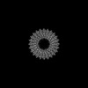 | |||||||||
|---|---|---|---|---|---|---|---|---|---|---|
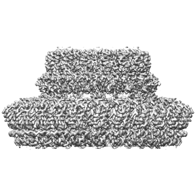
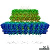
| Title | Inner membrane ring and secretin N0 N1 domains of the type 3 secretion system of Shigella flexneri | |||||||||
 Map data Map data | Main map sharpened and masked | |||||||||
 Sample Sample |
| |||||||||
 Keywords Keywords | type 3 secretion / inner membrane ring / secretin / shigella / infection / PROTEIN TRANSPORT | |||||||||
| Function / homology |  Function and homology information Function and homology informationtype III protein secretion system complex / type II protein secretion system complex / protein secretion by the type III secretion system / protein secretion / cell outer membrane / plasma membrane Similarity search - Function | |||||||||
| Biological species |  Shigella flexneri (bacteria) Shigella flexneri (bacteria) | |||||||||
| Method | single particle reconstruction / cryo EM / Resolution: 3.34 Å | |||||||||
 Authors Authors | Lunelli M | |||||||||
| Funding support |  Germany, 1 items Germany, 1 items
| |||||||||
 Citation Citation | Journal: Protein Sci / Year: 2023 Title: Integrative structural analysis of the type III secretion system needle complex from Shigella flexneri. Authors: Lara Flacht / Michele Lunelli / Karol Kaszuba / Zhuo Angel Chen / Francis J O' Reilly / Juri Rappsilber / Jan Kosinski / Michael Kolbe /  Abstract: The type III secretion system (T3SS) is a large, transmembrane protein machinery used by various pathogenic gram-negative bacteria to transport virulence factors into the host cell during infection. ...The type III secretion system (T3SS) is a large, transmembrane protein machinery used by various pathogenic gram-negative bacteria to transport virulence factors into the host cell during infection. Understanding the structure of T3SSs is crucial for future developments of therapeutics that could target this system. However, much of the knowledge about the structure of T3SS is available only for Salmonella, and it is unclear how this large assembly is conserved across species. Here, we combined cryo-electron microscopy, cross-linking mass spectrometry, and integrative modeling to determine the structure of the T3SS needle complex from Shigella flexneri. We show that the Shigella T3SS exhibits unique features distinguishing it from other structurally characterized T3SSs. The secretin pore complex adopts a new fold of its C-terminal S domain and the pilotin MxiM[SctG] locates around the outer surface of the pore. The export apparatus structure exhibits a conserved pseudohelical arrangement but includes the N-terminal domain of the SpaS[SctU] subunit, which was not present in any of the previously published virulence-related T3SS structures. Similar to other T3SSs, however, the apparatus is anchored within the needle complex by a network of flexible linkers that either adjust conformation to connect to equivalent patches on the secretin oligomer or bind distinct surface patches at the same height of the export apparatus. The conserved and unique features delineated by our analysis highlight the necessity to analyze T3SS in a species-specific manner, in order to fully understand the underlying molecular mechanisms of these systems. The structure of the type III secretion system from Shigella flexneri delineates conserved and unique features, which could be used for the development of broad-range therapeutics. | |||||||||
| History |
|
- Structure visualization
| Supplemental images |
|---|
- Downloads & links
-EMDB archive
| Map data | emd_15702.map.gz | 28.8 MB | EMDB map data format | |
|---|---|---|---|---|
| Header (meta data) | emd-15702-v30.xmlemd-15702.xml | 20.5 KB 20.5 KB | Display Display | EMDB header |
| FSC (resolution estimation) | emd_15702_fsc.xml | 16.9 KB | Display | FSC data file |
| Images |  emd_15702.png emd_15702.png | 114.2 KB | ||
| Masks | emd_15702_msk_1.map | 421.9 MB | Mask map | |
| Filedesc metadata | emd-15702.cif.gz | 6.3 KB | ||
| Others | emd_15702_half_map_1.map.gzemd_15702_half_map_2.map.gz | 329.2 MB 329.2 MB | ||
| Archive directory |  http://ftp.pdbj.org/pub/emdb/structures/EMD-15702ftp://ftp.pdbj.org/pub/emdb/structures/EMD-15702 http://ftp.pdbj.org/pub/emdb/structures/EMD-15702ftp://ftp.pdbj.org/pub/emdb/structures/EMD-15702 | HTTPS FTP |
-Related structure data
| Related structure data |  8axnMC 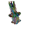 8axkC  8axlC M: atomic model generated by this map C: citing same article ( |
|---|---|
| Similar structure data |


-Links
| EMDB pages | EMDB (EBI/PDBe) / EMDataResource |
|---|---|
| Related items in Molecule of the Month |


-Map
| File | Download / File: emd_15702.map.gz / Format: CCP4 / Size: 421.9 MB / Type: IMAGE STORED AS FLOATING POINT NUMBER (4 BYTES) | ||||||||||||||||||||||||||||||||||||
|---|---|---|---|---|---|---|---|---|---|---|---|---|---|---|---|---|---|---|---|---|---|---|---|---|---|---|---|---|---|---|---|---|---|---|---|---|---|
| Annotation | Main map sharpened and masked | ||||||||||||||||||||||||||||||||||||
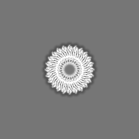
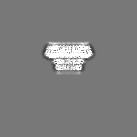
| Projections & slices | Image control
Images are generated by Spider. | ||||||||||||||||||||||||||||||||||||
| Voxel size | X=Y=Z: 1.38369 Å | ||||||||||||||||||||||||||||||||||||
| Density |
| ||||||||||||||||||||||||||||||||||||
| Symmetry | Space group: 1 | ||||||||||||||||||||||||||||||||||||
| Details | EMDB XML:
|
 Z (Sec.)
 Y (Row.)
 X (Col.)


-Supplemental data
-Mask #1
| File | emd_15702_msk_1.map | ||||||||||||
|---|---|---|---|---|---|---|---|---|---|---|---|---|---|
| Projections & Slices |
| ||||||||||||
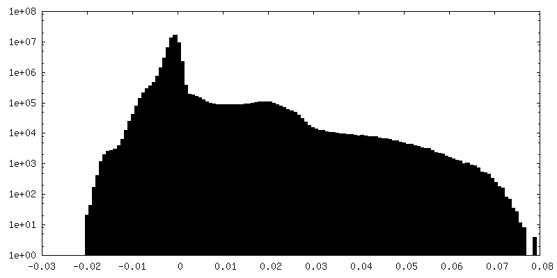
| Density Histograms |


-Half map: #1
| File | emd_15702_half_map_1.map | ||||||||||||
|---|---|---|---|---|---|---|---|---|---|---|---|---|---|
| Projections & Slices |
| ||||||||||||
| Density Histograms |


-Half map: #2
| File | emd_15702_half_map_2.map | ||||||||||||
|---|---|---|---|---|---|---|---|---|---|---|---|---|---|
| Projections & Slices |
| ||||||||||||
| Density Histograms |


- Sample components
-Entire : Needle complex of the type 3 secretion system
| Entire | Name: Needle complex of the type 3 secretion system |
|---|---|
| Components |
|
-Supramolecule #1: Needle complex of the type 3 secretion system
| Supramolecule | Name: Needle complex of the type 3 secretion system / type: complex / ID: 1 / Parent: 0 / Macromolecule list: all |
|---|---|
| Source (natural) | Organism: Shigella flexneri (bacteria) |
-Supramolecule #2: Inner membrane ring
| Supramolecule | Name: Inner membrane ring / type: complex / ID: 2 / Parent: 1 / Macromolecule list: #1-#2 |
|---|---|
| Source (natural) | Organism: Shigella flexneri (bacteria) |
-Supramolecule #3: Connector
| Supramolecule | Name: Connector / type: complex / ID: 3 / Parent: 1 / Macromolecule list: #3 Details: Ring formed by the domains N0 and N1 of the secretin MxiD |
|---|---|
| Source (natural) | Organism: Shigella flexneri (bacteria) |
-Macromolecule #1: Protein MxiG
| Macromolecule | Name: Protein MxiG / type: protein_or_peptide / ID: 1 / Number of copies: 24 / Enantiomer: LEVO |
|---|---|
| Source (natural) | Organism: Shigella flexneri (bacteria) |
| Molecular weight | Theoretical: 43.053844 KDa |
| Sequence | String: MSEAKNSNLA PFRLLVKLTN GVGDEFPLYY GNNLIVLGRT IETLEFGNDN FPENIIPVTD SKSDGIIYLT ISKDNICQFS DEKGEQIDI NSQFNSFEYD GISFHLKNMR EDKSRGHILN GMYKNHSVFF FFAVIVVLII IFSLSLKKDE VKEIAEIIDD K RYGIVNTG ...String: MSEAKNSNLA PFRLLVKLTN GVGDEFPLYY GNNLIVLGRT IETLEFGNDN FPENIIPVTD SKSDGIIYLT ISKDNICQFS DEKGEQIDI NSQFNSFEYD GISFHLKNMR EDKSRGHILN GMYKNHSVFF FFAVIVVLII IFSLSLKKDE VKEIAEIIDD K RYGIVNTG QCNYILAETQ NDAVWASVAL NKTGFTKCRY ILVSNKEINR IQQYINQRFP FINLYVLNLV SDKAELLVFL SK ERNSSKD TELDKLKNAL IVEFPYIKNI KFNYLSDHNA RGDAKGIFTK VNVQYKEICE NNKVTYSVRE ELTDEKLELI NRL ISEHKN IYGDQYIEFS VLLIDDDFKG KSYLNSKDSY VMLNDKHWFF LDKNK UniProtKB: Protein MxiG |
-Macromolecule #2: Lipoprotein MxiJ
| Macromolecule | Name: Lipoprotein MxiJ / type: protein_or_peptide / ID: 2 / Number of copies: 24 / Enantiomer: LEVO |
|---|---|
| Source (natural) | Organism: Shigella flexneri (bacteria) |
| Molecular weight | Theoretical: 27.542055 KDa |
| Sequence | String: MIRYKGFILF LLLMLIGCEQ REELISNLSQ RQANEIISVL ERHNITARKV DGGKQGISVQ VEKGTFASAV DLMRMYDLPN PERVDISQM FPTDSLVSSP RAEKARLYSA IEQRLEQSLV SIGGVISAKI HVSYDLEEKN ISSKPMHISV IAIYDSPKES E LLVSNIKR ...String: MIRYKGFILF LLLMLIGCEQ REELISNLSQ RQANEIISVL ERHNITARKV DGGKQGISVQ VEKGTFASAV DLMRMYDLPN PERVDISQM FPTDSLVSSP RAEKARLYSA IEQRLEQSLV SIGGVISAKI HVSYDLEEKN ISSKPMHISV IAIYDSPKES E LLVSNIKR FLKNTFSDVK YENISVILTP KEEYVYTNVQ PVKEVKSEFL TNEVIYLFLG MAVLVVILLV WAFKTGWFKR NK I UniProtKB: Lipoprotein MxiJ |
-Macromolecule #3: Outer membrane protein MxiD
| Macromolecule | Name: Outer membrane protein MxiD / type: protein_or_peptide / ID: 3 / Number of copies: 16 / Enantiomer: LEVO |
|---|---|
| Source (natural) | Organism: Shigella flexneri (bacteria) |
| Molecular weight | Theoretical: 63.230414 KDa |
| Sequence | String: MKKFNIKSLT LLIVLLPLIV NANNIDSHLL EQNDIAKYVA QSDTVGSFFE RFSALLNYPI VVSKQAAKKR ISGEFDLSNP EEMLEKLTL LVGLIWYKDG NALYIYDSGE LISKVILLEN ISLNYLIQYL KDANLYDHRY PIRGNISDKT FYISGPPALV E LVANTATL ...String: MKKFNIKSLT LLIVLLPLIV NANNIDSHLL EQNDIAKYVA QSDTVGSFFE RFSALLNYPI VVSKQAAKKR ISGEFDLSNP EEMLEKLTL LVGLIWYKDG NALYIYDSGE LISKVILLEN ISLNYLIQYL KDANLYDHRY PIRGNISDKT FYISGPPALV E LVANTATL LDKQVSSIGT DKVNFGVIKL KNTFVSDRTY NMRGEDIVIP GVATVVERLL NNGKALSNRQ AQNDPMPPFN IT QKVSEDS NDFSFSSVTN SSILEDVSLI AYPETNSILV KGNDQQIQII RDIITQLDVA KRHIELSLWI IDIDKSELNN LGV NWQGTA SFGDSFGASF NMSSSASIST LDGNKFIASV MALNQKKKAN VVSRPVILTQ ENIPAIFDNN RTFYVSLVGE RNSS LEHVT YGTLINVIPR FSSRGQIEMS LTIEDGTGNS QSNYNYNNEN TSVLPEVGRT KISTIARVPQ GKSLLIGGYT HETNS NEII SIPFLSSIPV IGNVFKYKTS NISNIVRVFL IQPREIKESS YYNTAEYKSL ISEREIQKTT QIIPSETTLL EDEKSL VSY LNY UniProtKB: Type 3 secretion system secretin |
-Experimental details
-Structure determination
| Method | cryo EM |
|---|---|
 Processing Processing | single particle reconstruction |
| Aggregation state | particle |
-Sample preparation
| Buffer | pH: 8 |
|---|---|
| Grid | Model: Quantifoil R2/1 / Material: COPPER / Pretreatment - Type: GLOW DISCHARGE / Pretreatment - Atmosphere: AIR |
| Vitrification | Cryogen name: ETHANE / Chamber humidity: 100 % / Chamber temperature: 295 K / Instrument: FEI VITROBOT MARK IV |
- Electron microscopy
| Microscope | FEI TITAN KRIOS |
|---|---|
| Image recording | Film or detector model: FEI FALCON II (4k x 4k) / Detector mode: INTEGRATING / Digitization - Dimensions - Width: 4096 pixel / Digitization - Dimensions - Height: 4096 pixel / Digitization - Frames/image: 1-6 / Number grids imaged: 1 / Number real images: 5238 / Average exposure time: 1.5 sec. / Average electron dose: 25.0 e/Å2 |
| Electron beam | Acceleration voltage: 300 kV / Electron source:  FIELD EMISSION GUN FIELD EMISSION GUN |
| Electron optics | Illumination mode: OTHER / Imaging mode: BRIGHT FIELD / Cs: 2.7 mm / Nominal defocus max: 4.0 µm / Nominal defocus min: 1.5 µm / Nominal magnification: 101179 |
| Sample stage | Specimen holder model: FEI TITAN KRIOS AUTOGRID HOLDER / Cooling holder cryogen: NITROGEN |
| Experimental equipment |  Model: Titan Krios / Image courtesy: FEI Company |