- EMDB-15700: Type 3 secretion system needle complex of Shigella flexneri -
+
Open data
ID or keywords:
Loading...
-
Basic information
Entry
Database: EMDB / ID: EMD-15700
Title
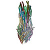
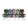
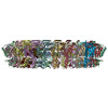
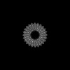
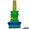
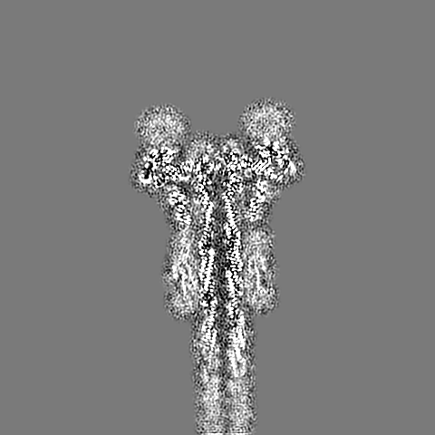
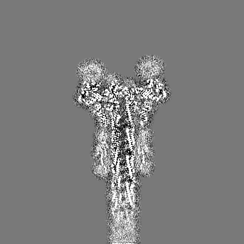
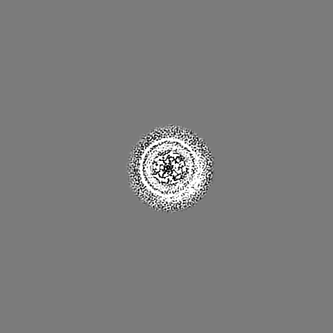
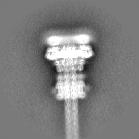
Type 3 secretion system needle complex of Shigella flexneri
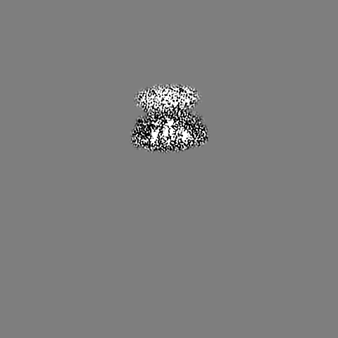
Map data
Main map (sharpened and masked)
Sample
Complex: Needle complex of the type 3 secretion system
Complex: Export apparatus core
Protein or peptide: Surface presentation of antigens protein SpaP
Protein or peptide: Surface presentation of antigens protein SpaR
Protein or peptide: Surface presentation of antigens protein SpaQ
Protein or peptide: Surface presentation of antigens protein SpaS
Complex: Inner rod
Protein or peptide: Protein MxiI
Complex: Needle filament
Protein or peptide: Protein MxiH
Complex: Ring formed by the N1 domain of the secretin MxiD
Protein or peptide: Outer membrane protein MxiD
Complex: MxiJ loop interacting with the export apparatus
Protein or peptide: Lipoprotein MxiJ
Keywords
type 3 secretion / needle complex / virulence factor / shigella / infection / PROTEIN TRANSPORT
Function / homology
Function and homology information
type III protein secretion system complex / type II protein secretion system complex / protein secretion by the type III secretion system / protein secretion / protein targeting / cell outer membrane / protein transport / cell surface / extracellular region / identical protein binding / plasma membrane Similarity search - Function
: / Type III secretion protein SpaR/YscT / Type III secretion protein HrpO / Yop virulence translocation protein R / Type III exporter system, secretion apparatus protein BsaZ / Type III secretion system lipoprotein HrcJ/YscJ / Type III secretion system substrate exporter / Type III secretion system substrate exporter, C-terminal / FlhB HrpN YscU SpaS Family / : ...: / Type III secretion protein SpaR/YscT / Type III secretion protein HrpO / Yop virulence translocation protein R / Type III exporter system, secretion apparatus protein BsaZ / Type III secretion system lipoprotein HrcJ/YscJ / Type III secretion system substrate exporter / Type III secretion system substrate exporter, C-terminal / FlhB HrpN YscU SpaS Family / : / SPI-1 type 3 secretion system secretin, N0 domain / Type III secretion system outer membrane pore YscC/HrcC / Type III secretion, needle-protein-like / Type III secretion, needle-protein-like superfamily / Type III secretion needle MxiH, YscF, SsaG, EprI, PscF, EscF / Type III secretion system, needle protein / Bacterial export protein family 3 / Bacterial export proteins, family 3 / Type III secretion system inner membrane R protein / Bacterial export proteins, family 1 / Flagella transport protein fliP family signature 1. / Type III secretion system inner membrane P protein / FliP family / Flagella transport protein fliP family signature 2. / : / Bacterial type II secretion system protein D signature. / Type II secretion system protein GspD, conserved site / NolW-like / NolW-like superfamily / Bacterial type II/III secretion system short domain / Type II/III secretion system / Bacterial type II and III secretion system protein / Lipoprotein YscJ/Flagellar M-ring protein / Secretory protein of YscJ/FliF RBM domain / Flagellar M-ring , N-terminal / AMP-binding enzyme, C-terminal domain superfamily / Prokaryotic membrane lipoprotein lipid attachment site profile. Similarity search - Domain/homology
Surface presentation of antigens protein SpaP / Surface presentation of antigens protein SpaQ / Surface presentation of antigens protein SpaR / Surface presentation of antigens protein SpaS / Type 3 secretion system needle filament protein / Protein MxiI / Type 3 secretion system secretin / Lipoprotein MxiJ Similarity search - Component
Biological species
Shigella flexneri (bacteria)
Method
single particle reconstruction / cryo EM / Resolution: 4.05 Å
Journal: Protein Sci / Year: 2023 Title: Integrative structural analysis of the type III secretion system needle complex from Shigella flexneri. Authors: Lara Flacht / Michele Lunelli / Karol Kaszuba / Zhuo Angel Chen / Francis J O' Reilly / Juri Rappsilber / Jan Kosinski / Michael Kolbe / Abstract: The type III secretion system (T3SS) is a large, transmembrane protein machinery used by various pathogenic gram-negative bacteria to transport virulence factors into the host cell during infection. ...The type III secretion system (T3SS) is a large, transmembrane protein machinery used by various pathogenic gram-negative bacteria to transport virulence factors into the host cell during infection. Understanding the structure of T3SSs is crucial for future developments of therapeutics that could target this system. However, much of the knowledge about the structure of T3SS is available only for Salmonella, and it is unclear how this large assembly is conserved across species. Here, we combined cryo-electron microscopy, cross-linking mass spectrometry, and integrative modeling to determine the structure of the T3SS needle complex from Shigella flexneri. We show that the Shigella T3SS exhibits unique features distinguishing it from other structurally characterized T3SSs. The secretin pore complex adopts a new fold of its C-terminal S domain and the pilotin MxiM[SctG] locates around the outer surface of the pore. The export apparatus structure exhibits a conserved pseudohelical arrangement but includes the N-terminal domain of the SpaS[SctU] subunit, which was not present in any of the previously published virulence-related T3SS structures. Similar to other T3SSs, however, the apparatus is anchored within the needle complex by a network of flexible linkers that either adjust conformation to connect to equivalent patches on the secretin oligomer or bind distinct surface patches at the same height of the export apparatus. The conserved and unique features delineated by our analysis highlight the necessity to analyze T3SS in a species-specific manner, in order to fully understand the underlying molecular mechanisms of these systems. The structure of the type III secretion system from Shigella flexneri delineates conserved and unique features, which could be used for the development of broad-range therapeutics.
In the structure databanks used in Yorodumi, some data are registered as the other names, "COVID-19 virus" and "2019-nCoV". Here are the details of the virus and the list of structure data.
Jan 31, 2019. EMDB accession codes are about to change! (news from PDBe EMDB page)
EMDB accession codes are about to change! (news from PDBe EMDB page)
The allocation of 4 digits for EMDB accession codes will soon come to an end. Whilst these codes will remain in use, new EMDB accession codes will include an additional digit and will expand incrementally as the available range of codes is exhausted. The current 4-digit format prefixed with “EMD-” (i.e. EMD-XXXX) will advance to a 5-digit format (i.e. EMD-XXXXX), and so on. It is currently estimated that the 4-digit codes will be depleted around Spring 2019, at which point the 5-digit format will come into force.
The EM Navigator/Yorodumi systems omit the EMD- prefix.
Related info.:Q: What is EMD? / ID/Accession-code notation in Yorodumi/EM Navigator
Yorodumi is a browser for structure data from EMDB, PDB, SASBDB, etc.
This page is also the successor to EM Navigator detail page, and also detail information page/front-end page for Omokage search.
The word "yorodu" (or yorozu) is an old Japanese word meaning "ten thousand". "mi" (miru) is to see.
Related info.:EMDB / PDB / SASBDB / Comparison of 3 databanks / Yorodumi Search / Aug 31, 2016. New EM Navigator & Yorodumi / Yorodumi Papers / Jmol/JSmol / Function and homology information / Changes in new EM Navigator and Yorodumi
 Movie
Movie Controller


 Open data
 Basic information
 Map data
 Sample
 Keywords
 Function and homology information
 Shigella flexneri (bacteria)
 Authors
 Germany, 1 items
  Citation
 Structure visualization
 Downloads & links
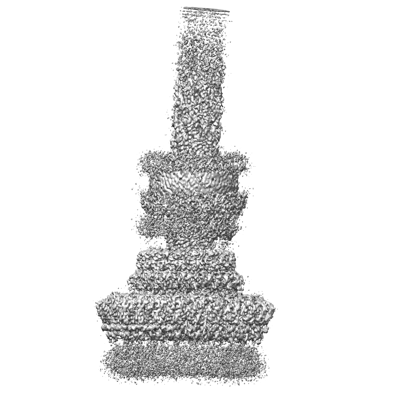 emd_15700.png
 http://ftp.pdbj.org/pub/emdb/structures/EMD-15700
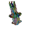


 Z (Sec.)
 Y (Row.)
 X (Col.)


 Sample components
 Processing
 Electron microscopy
 FIELD EMISSION GUN