 Movie
Movie Controller

[English] 日本語
 Yorodumi
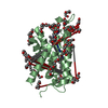
- PDB-7vw1: Structure of a dimeric periplasmic protein bound with cuprous ions -
+ Open data

- Basic information
| Entry | Database: PDB / ID: 7vw1 | ||||||
|---|---|---|---|---|---|---|---|
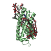
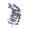
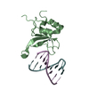
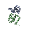
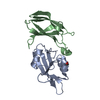
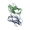
| Title | Structure of a dimeric periplasmic protein bound with cuprous ions | ||||||
 Components Components | DUF305 domain-containing protein | ||||||
 Keywords Keywords | METAL BINDING PROTEIN / Copper binding protein / Metal homeostasis | ||||||
| Function / homology | Domain of unknown function DUF305, CopM-like / Domain of unknown function (DUF305) / Ferritin-like / COPPER (I) ION / DUF305 domain-containing protein Function and homology information Function and homology information | ||||||
| Biological species |  | ||||||
| Method |  X-RAY DIFFRACTION / SYNCHROTRON / MOLECULAR REPLACEMENT / Resolution: 2.494 Å X-RAY DIFFRACTION / SYNCHROTRON / MOLECULAR REPLACEMENT / Resolution: 2.494 Å | ||||||
 Authors Authors | Yang, J. / Liu, L. | ||||||
| Funding support |  China, 1items China, 1items
| ||||||
 Citation Citation | Journal: J.Inorg.Biochem. / Year: 2022 Title: Structural basis of copper binding by a dimeric periplasmic protein forming a six-helical bundle. Authors: Yang, J. / Gao, M. / Wang, J. / He, C. / Wang, X. / Liu, L. | ||||||
| History |
|
- Structure visualization
| Structure viewer | Molecule: MolmilJmol/JSmol |
|---|
- Downloads & links
-Download
| PDBx/mmCIF format | 7vw1.cif.gz | 86.7 KB | Display | PDBx/mmCIF format |
|---|---|---|---|---|
| PDB format | pdb7vw1.ent.gz | 64.1 KB | Display | PDB format |
| PDBx/mmJSON format | 7vw1.json.gz | Tree view | PDBx/mmJSON format | |
| Others |  Other downloads Other downloads |
-Validation report
| Arichive directory | https://data.pdbj.org/pub/pdb/validation_reports/vw/7vw1ftp://data.pdbj.org/pub/pdb/validation_reports/vw/7vw1 | HTTPS FTP |
|---|
-Related structure data
| Related structure data | 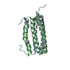 7vw0C 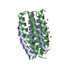 7vw2C  5ffbS S: Starting model for refinement C: citing same article ( |
|---|---|
| Similar structure data |


-Links
 PDBj
PDBj


- Assembly
| Deposited unit | 
| |||||||||||||||||||||||||||
|---|---|---|---|---|---|---|---|---|---|---|---|---|---|---|---|---|---|---|---|---|---|---|---|---|---|---|---|---|
| 1 |
| |||||||||||||||||||||||||||
| Unit cell |
| |||||||||||||||||||||||||||
| Noncrystallographic symmetry (NCS) | NCS domain:
NCS domain segments:
|
-Components
| #1: Protein | Mass: 10887.481 Da / Num. of mol.: 2 Source method: isolated from a genetically manipulated source Source: (gene. exp.) #2: Chemical | ChemComp-CU1 /   Mass: 63.546 Da / Num. of mol.: 5 / Source method: obtained synthetically / Formula: Cu / Feature type: SUBJECT OF INVESTIGATION Mass: 63.546 Da / Num. of mol.: 5 / Source method: obtained synthetically / Formula: Cu / Feature type: SUBJECT OF INVESTIGATION#3: Water | ChemComp-HOH / |  Mass: 18.015 Da / Num. of mol.: 38 / Source method: isolated from a natural source / Formula: H2O Mass: 18.015 Da / Num. of mol.: 38 / Source method: isolated from a natural source / Formula: H2OHas ligand of interest | Y | |
|---|
-Experimental details
-Experiment
| Experiment | Method: X-RAY DIFFRACTION / Number of used crystals: 1 |
|---|
- Sample preparation
| Crystal | Density Matthews: 3.1 Å3/Da / Density % sol: 60.22 % |
|---|---|
| Crystal grow | Temperature: 289 K / Method: vapor diffusion, sitting drop / Details: Lithium chloride, AmSO4 |
-Data collection
| Diffraction | Mean temperature: 100 K / Serial crystal experiment: N |
|---|---|
| Diffraction source | Source: SYNCHROTRON / Site: SSRF / Beamline: BL19U1 / Wavelength: 0.9792 Å |
| Detector | Type: DECTRIS PILATUS 6M / Detector: PIXEL / Date: Oct 1, 2021 |
| Radiation | Protocol: SINGLE WAVELENGTH / Monochromatic (M) / Laue (L): M / Scattering type: x-ray |
| Radiation wavelength | Wavelength: 0.9792 Å / Relative weight: 1 |
| Reflection | Resolution: 2.494→50 Å / Num. obs: 9526 / % possible obs: 99.9 % / Redundancy: 19.3 % / Biso Wilson estimate: 39.39 Å2 / CC1/2: 0.983 / Rmerge(I) obs: 0.132 / Rpim(I) all: 0.049 / Net I/σ(I): 19.3 |
| Reflection shell | Resolution: 2.494→2.59 Å / Rmerge(I) obs: 0.905 / Num. unique obs: 930 / CC1/2: 0.956 / Rpim(I) all: 0.309 |
- Processing
| Software |
| ||||||||||||||||||||||||||||||||||||||||||
|---|---|---|---|---|---|---|---|---|---|---|---|---|---|---|---|---|---|---|---|---|---|---|---|---|---|---|---|---|---|---|---|---|---|---|---|---|---|---|---|---|---|---|---|
| Refinement | Method to determine structure: MOLECULAR REPLACEMENT Starting model: 5FFB Resolution: 2.494→34.621 Å / SU ML: 0.38 / Cross valid method: THROUGHOUT / σ(F): 1.37 / Phase error: 29.12 / Stereochemistry target values: ML
| ||||||||||||||||||||||||||||||||||||||||||
| Solvent computation | Shrinkage radii: 0.9 Å / VDW probe radii: 1.11 Å / Solvent model: FLAT BULK SOLVENT MODEL | ||||||||||||||||||||||||||||||||||||||||||
| Displacement parameters | Biso max: 111.37 Å2 / Biso mean: 52.2696 Å2 / Biso min: 30.33 Å2 | ||||||||||||||||||||||||||||||||||||||||||
| Refinement step | Cycle: final / Resolution: 2.494→34.621 Å
| ||||||||||||||||||||||||||||||||||||||||||
| Refine LS restraints |
| ||||||||||||||||||||||||||||||||||||||||||
| Refine LS restraints NCS |
| ||||||||||||||||||||||||||||||||||||||||||
| LS refinement shell | Refine-ID: X-RAY DIFFRACTION / Rfactor Rfree error: 0
| ||||||||||||||||||||||||||||||||||||||||||
| Refinement TLS params. | Method: refined / Origin x: 1.435 Å / Origin y: 23.7456 Å / Origin z: -3.4004 Å
| ||||||||||||||||||||||||||||||||||||||||||
| Refinement TLS group |
|
