[English] 日本語
Yorodumi
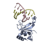
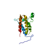
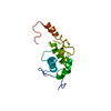
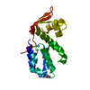
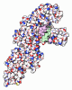
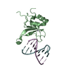
- PDB-3odc: Human PARP-1 zinc finger 2 (Zn2) bound to DNA -

+
Open data


ID or keywords:

Loading...

-
Basic information

Entry
Database: PDB / ID: 3odc
TitleHuman PARP-1 zinc finger 2 (Zn2) bound to DNA
Components
  • 5'-D(*CP*CP*CP*AP*GP*AP*CP*G)-3'
  • 5'-D(*CP*GP*TP*CP*TP*GP*GP*G)-3'
  • Poly [ADP-ribose] polymerase 1
KeywordsDNA BINDING PROTEIN/DNA / protein-DNA complex / PARP zinc finger / DNA BINDING PROTEIN-DNA complex
Function / homology
Function and homology information


NAD+-histone H2BS6 serine ADP-ribosyltransferase activity / NAD+-histone H3S10 serine ADP-ribosyltransferase activity / NAD+-histone H2BE35 glutamate ADP-ribosyltransferase activity / positive regulation of myofibroblast differentiation / negative regulation of ATP biosynthetic process / NAD+-protein-tyrosine ADP-ribosyltransferase activity / NAD+-protein-histidine ADP-ribosyltransferase activity / non-sequence-specific DNA binding, bending / regulation of base-excision repair / mitochondrial DNA metabolic process ...NAD+-histone H2BS6 serine ADP-ribosyltransferase activity / NAD+-histone H3S10 serine ADP-ribosyltransferase activity / NAD+-histone H2BE35 glutamate ADP-ribosyltransferase activity / positive regulation of myofibroblast differentiation / negative regulation of ATP biosynthetic process / NAD+-protein-tyrosine ADP-ribosyltransferase activity / NAD+-protein-histidine ADP-ribosyltransferase activity / non-sequence-specific DNA binding, bending / regulation of base-excision repair / mitochondrial DNA metabolic process / regulation of circadian sleep/wake cycle, non-REM sleep / vRNA Synthesis / carbohydrate biosynthetic process / NAD+-protein-serine ADP-ribosyltransferase activity / NAD DNA ADP-ribosyltransferase activity / single-strand break-containing DNA binding / mitochondrial DNA repair / DNA ADP-ribosylation / regulation of oxidative stress-induced neuron intrinsic apoptotic signaling pathway / signal transduction involved in regulation of gene expression / ATP generation from poly-ADP-D-ribose / replication fork reversal / positive regulation of necroptotic process / establishment of protein localization to chromatin / positive regulation of intracellular estrogen receptor signaling pathway / transcription regulator activator activity / response to aldosterone / HDR through MMEJ (alt-NHEJ) / single strand break repair / positive regulation of DNA-templated transcription, elongation / NAD+ ADP-ribosyltransferase / protein auto-ADP-ribosylation / negative regulation of telomere maintenance via telomere lengthening / cellular response to zinc ion / NAD+-protein-aspartate ADP-ribosyltransferase activity / protein poly-ADP-ribosylation / NAD+-protein-glutamate ADP-ribosyltransferase activity / negative regulation of cGAS/STING signaling pathway / positive regulation of cardiac muscle hypertrophy / negative regulation of transcription elongation by RNA polymerase II / decidualization / NAD+-protein mono-ADP-ribosyltransferase activity / positive regulation of mitochondrial depolarization / protein autoprocessing / macrophage differentiation / R-SMAD binding / negative regulation of adipose tissue development / nuclear replication fork / Transferases; Glycosyltransferases; Pentosyltransferases / positive regulation of SMAD protein signal transduction / POLB-Dependent Long Patch Base Excision Repair / NAD+ poly-ADP-ribosyltransferase activity / transforming growth factor beta receptor signaling pathway / SUMOylation of DNA damage response and repair proteins / positive regulation of double-strand break repair via homologous recombination / nucleosome binding / protein localization to chromatin / nucleotidyltransferase activity / response to gamma radiation / negative regulation of innate immune response / telomere maintenance / nuclear estrogen receptor binding / protein modification process / site of DNA damage / mitochondrion organization / cellular response to nerve growth factor stimulus / Downregulation of SMAD2/3:SMAD4 transcriptional activity / positive regulation of protein localization to nucleus / protein-DNA complex / fibrillar center / transcription by RNA polymerase II / DNA Damage Recognition in GG-NER / NAD binding / cellular response to amyloid-beta / enzyme activator activity / histone deacetylase binding / Dual Incision in GG-NER / Formation of Incision Complex in GG-NER / cellular response to UV / double-strand break repair / cellular response to insulin stimulus / nuclear envelope / regulation of protein localization / site of double-strand break / transcription regulator complex / cellular response to oxidative stress / damaged DNA binding / response to ethanol / RNA polymerase II-specific DNA-binding transcription factor binding / nuclear body / positive regulation of canonical NF-kappaB signal transduction / chromosome, telomeric region / innate immune response / negative regulation of DNA-templated transcription / ubiquitin protein ligase binding / DNA repair / apoptotic process / chromatin binding / DNA damage response / nucleolus
Similarity search - Function
Zinc finger, PARP-type / first zn-finger domain of poly(adp-ribose) polymerase-1 / Poly [ADP-ribose] polymerase / PADR1 domain / PADR1 domain superfamily / : / PADR1 domain, zinc ribbon fold / PADR1, N-terminal helical domain / PADR1 domain profile. / PADR1 ...Zinc finger, PARP-type / first zn-finger domain of poly(adp-ribose) polymerase-1 / Poly [ADP-ribose] polymerase / PADR1 domain / PADR1 domain superfamily / : / PADR1 domain, zinc ribbon fold / PADR1, N-terminal helical domain / PADR1 domain profile. / PADR1 / Zinc finger poly(ADP-ribose) polymerase (PARP)-type signature. / Zinc finger, PARP-type superfamily / Poly(ADP-ribose) polymerase and DNA-Ligase Zn-finger region / Zinc finger poly(ADP-ribose) polymerase (PARP)-type profile. / Poly(ADP-ribose) polymerase and DNA-Ligase Zn-finger region / Zinc finger, PARP-type / : / Poly(ADP-ribose) polymerase, regulatory domain / WGR domain / WGR domain superfamily / WGR domain / WGR domain profile. / Proposed nucleic acid binding domain / Poly(ADP-ribose) polymerase, regulatory domain superfamily / Poly(ADP-ribose) polymerase, regulatory domain / PARP alpha-helical domain profile. / BRCA1 C Terminus (BRCT) domain / Poly(ADP-ribose) polymerase catalytic domain / Poly(ADP-ribose) polymerase, catalytic domain / PARP catalytic domain profile. / breast cancer carboxy-terminal domain / BRCT domain profile. / BRCT domain / BRCT domain superfamily / 2-Layer Sandwich / Alpha Beta
Similarity search - Domain/homology
DNA / Poly [ADP-ribose] polymerase 1
Similarity search - Component
Biological speciesHomo sapiens (human)
MethodX-RAY DIFFRACTION / SYNCHROTRON / SAD / Resolution: 2.8 Å
AuthorsPascal, J.M. / Langelier, M.-F.
CitationJournal: J.Biol.Chem. / Year: 2011
Title: Crystal Structures of Poly(ADP-ribose) Polymerase-1 (PARP-1) Zinc Fingers Bound to DNA: STRUCTURAL AND FUNCTIONAL INSIGHTS INTO DNA-DEPENDENT PARP-1 ACTIVITY.
Authors: Langelier, M.F. / Planck, J.L. / Roy, S. / Pascal, J.M.
History
DepositionAug 11, 2010Deposition site: RCSB / Processing site: RCSB
Revision 1.0Jan 12, 2011Provider: repository / Type: Initial release
Revision 1.1Jul 13, 2011Group: Version format compliance
Revision 1.2Nov 8, 2017Group: Refinement description / Category: software
Item: _software.classification / _software.contact_author ..._software.classification / _software.contact_author / _software.contact_author_email / _software.date / _software.language / _software.location / _software.name / _software.type / _software.version
Revision 1.3Feb 21, 2024Group: Data collection / Database references / Derived calculations
Category: chem_comp_atom / chem_comp_bond ...chem_comp_atom / chem_comp_bond / database_2 / pdbx_struct_conn_angle / struct_conn / struct_ref_seq_dif / struct_site
Item: _database_2.pdbx_DOI / _database_2.pdbx_database_accession ..._database_2.pdbx_DOI / _database_2.pdbx_database_accession / _pdbx_struct_conn_angle.ptnr1_auth_asym_id / _pdbx_struct_conn_angle.ptnr1_auth_comp_id / _pdbx_struct_conn_angle.ptnr1_auth_seq_id / _pdbx_struct_conn_angle.ptnr1_label_asym_id / _pdbx_struct_conn_angle.ptnr1_label_atom_id / _pdbx_struct_conn_angle.ptnr1_label_comp_id / _pdbx_struct_conn_angle.ptnr1_label_seq_id / _pdbx_struct_conn_angle.ptnr2_auth_asym_id / _pdbx_struct_conn_angle.ptnr2_label_asym_id / _pdbx_struct_conn_angle.ptnr3_auth_asym_id / _pdbx_struct_conn_angle.ptnr3_auth_comp_id / _pdbx_struct_conn_angle.ptnr3_auth_seq_id / _pdbx_struct_conn_angle.ptnr3_label_asym_id / _pdbx_struct_conn_angle.ptnr3_label_atom_id / _pdbx_struct_conn_angle.ptnr3_label_comp_id / _pdbx_struct_conn_angle.ptnr3_label_seq_id / _pdbx_struct_conn_angle.value / _struct_conn.pdbx_dist_value / _struct_conn.ptnr1_auth_asym_id / _struct_conn.ptnr1_auth_comp_id / _struct_conn.ptnr1_auth_seq_id / _struct_conn.ptnr1_label_asym_id / _struct_conn.ptnr1_label_atom_id / _struct_conn.ptnr1_label_comp_id / _struct_conn.ptnr1_label_seq_id / _struct_conn.ptnr2_auth_asym_id / _struct_conn.ptnr2_label_asym_id / _struct_ref_seq_dif.details / _struct_site.pdbx_auth_asym_id / _struct_site.pdbx_auth_comp_id / _struct_site.pdbx_auth_seq_id

-
Structure visualization

Structure viewerMolecule:
MolmilJmol/JSmol

Downloads & links

-
Assembly

Deposited unit
A: Poly [ADP-ribose] polymerase 1
B: Poly [ADP-ribose] polymerase 1
C: 5'-D(*CP*CP*CP*AP*GP*AP*CP*G)-3'
D: 5'-D(*CP*GP*TP*CP*TP*GP*GP*G)-3'
E: 5'-D(*CP*CP*CP*AP*GP*AP*CP*G)-3'
F: 5'-D(*CP*GP*TP*CP*TP*GP*GP*G)-3'
hetero molecules


Theoretical massNumber of molelcules
Total (without water)35,2858
Polymers35,1546
Non-polymers1312
Water70339
1
A: Poly [ADP-ribose] polymerase 1
C: 5'-D(*CP*CP*CP*AP*GP*AP*CP*G)-3'
D: 5'-D(*CP*GP*TP*CP*TP*GP*GP*G)-3'
hetero molecules


Theoretical massNumber of molelcules
Total (without water)17,6424
Polymers17,5773
Non-polymers651
Water543
TypeNameSymmetry operationNumber
identity operation1_555x,y,z1
2
B: Poly [ADP-ribose] polymerase 1
E: 5'-D(*CP*CP*CP*AP*GP*AP*CP*G)-3'
F: 5'-D(*CP*GP*TP*CP*TP*GP*GP*G)-3'
hetero molecules


Theoretical massNumber of molelcules
Total (without water)17,6424
Polymers17,5773
Non-polymers651
Water543
TypeNameSymmetry operationNumber
identity operation1_555x,y,z1
Unit cell
Length a, b, c (Å)63.731, 63.731, 192.405
Angle α, β, γ (deg.)90.000, 90.000, 120.000
Int Tables number152
Space group name H-MP3121

-
Components

#1: Protein Poly [ADP-ribose] polymerase 1 / PARP-1 / NAD(+) ADP-ribosyltransferase 1 / ADPRT 1 / Poly[ADP-ribose] synthase 1


Mass: 12721.641 Da / Num. of mol.: 2 / Fragment: PARP-1 zinc finger 2, Zn2, UNP residues 105-206
Source method: isolated from a genetically manipulated source
Source: (gene. exp.) Homo sapiens (human) / Gene: ADPRT, PARP1, PPOL / Plasmid: pET24 / Production host: Escherichia coli (E. coli) / Strain (production host): BL21(DE3) Rosetta2 / References: UniProt: P09874, NAD+ ADP-ribosyltransferase
#2: DNA chain 5'-D(*CP*CP*CP*AP*GP*AP*CP*G)-3'


Mass: 2396.595 Da / Num. of mol.: 2 / Source method: obtained synthetically / Details: Chemically synthesized DNA
#3: DNA chain 5'-D(*CP*GP*TP*CP*TP*GP*GP*G)-3'


Mass: 2458.615 Da / Num. of mol.: 2 / Source method: obtained synthetically / Details: Chemically synthesized DNA
#4: Chemical ChemComp-ZN / ZINC ION


Mass: 65.409 Da / Num. of mol.: 2 / Source method: obtained synthetically / Formula: Zn
#5: Water ChemComp-HOH / water


Mass: 18.015 Da / Num. of mol.: 39 / Source method: isolated from a natural source / Formula: H2O

-
Experimental details

-
Experiment

ExperimentMethod: X-RAY DIFFRACTION / Number of used crystals: 1

-
Sample preparation

CrystalDensity Matthews: 3.21 Å3/Da / Density % sol: 61.67 %
Crystal growTemperature: 298 K / Method: vapor diffusion / pH: 6.5
Details: 1.36-1.42 M sodium citrate pH 6.5, vapor diffusion, temperature 298K

-
Data collection

DiffractionMean temperature: 100 K
Diffraction sourceSource: SYNCHROTRON / Site: NSLS / Beamline: X12C / Wavelength: 0.99 Å
DetectorType: ADSC QUANTUM 270 / Detector: CCD / Date: Sep 18, 2009
RadiationMonochromator: silicon(111) crystal / Protocol: SINGLE WAVELENGTH / Scattering type: x-ray
Radiation wavelengthWavelength: 0.99 Å / Relative weight: 1
ReflectionResolution: 2.8→50 Å / Num. obs: 11845 / % possible obs: 99.9 % / Redundancy: 8.1 % / Rmerge(I) obs: 0.092 / Χ2: 1 / Net I/σ(I): 8.3
Reflection shell
Resolution (Å)Redundancy (%)Rmerge(I) obsNum. unique allΧ2% possible all
2.8-2.858.20.7175710.982100
2.85-2.97.90.6256020.992100
2.9-2.968.30.6395520.986100
2.96-3.028.20.4595850.95100
3.02-3.088.20.3855821.02100
3.08-3.158.20.3085800.973100
3.15-3.238.20.2125720.979100
3.23-3.328.30.1635801.021100
3.32-3.428.20.1295711.034100
3.42-3.538.10.1135970.974100
3.53-3.658.30.15791.012100
3.65-3.88.20.0975900.991100
3.8-3.978.10.0875950.986100
3.97-4.188.30.0675731.025100
4.18-4.448.20.0635940.996100
4.44-4.798.10.066041.008100
4.79-5.278.10.0566041.03100
5.27-6.038.10.0546060.996100
6.03-7.597.90.0526221.027100
7.59-507.10.0366861.00898.7

-
Processing

Software
NameVersionClassificationNB
DENZOdata reduction
SCALEPACKdata scaling
PHENIX1.6.2_432refinement
PDB_EXTRACT3.1data extraction
CBASSdata collection
HKL-2000data reduction
SOLVEphasing
RefinementMethod to determine structure: SAD / Resolution: 2.8→36.262 Å / Occupancy max: 1 / Occupancy min: 0.5 / FOM work R set: 0.8113 / SU ML: 0.35 / σ(F): 1.35 / Stereochemistry target values: ML
RfactorNum. reflection% reflection
Rfree0.2398 556 4.71 %
Rwork0.1927 --
obs0.195 11798 99.93 %
Solvent computationShrinkage radii: 0.9 Å / VDW probe radii: 1.11 Å / Solvent model: FLAT BULK SOLVENT MODEL / Bsol: 34.698 Å2 / ksol: 0.337 e/Å3
Displacement parametersBiso max: 96.76 Å2 / Biso mean: 48.3666 Å2 / Biso min: 26.78 Å2
Baniso -1Baniso -2Baniso -3
1--0.5071 Å2-0 Å2-0 Å2
2---0.5071 Å20 Å2
3---1.0142 Å2
Refinement stepCycle: LAST / Resolution: 2.8→36.262 Å
ProteinNucleic acidLigandSolventTotal
Num. atoms1514 644 2 39 2199
Refine LS restraints
Refine-IDTypeDev idealNumber
X-RAY DIFFRACTIONf_bond_d0.012277
X-RAY DIFFRACTIONf_angle_d1.3053173
X-RAY DIFFRACTIONf_chiral_restr0.071337
X-RAY DIFFRACTIONf_plane_restr0.005300
X-RAY DIFFRACTIONf_dihedral_angle_d24.867917
LS refinement shell

Refine-ID: X-RAY DIFFRACTION / Total num. of bins used: 4 / % reflection obs: 100 %

Resolution (Å)Rfactor RfreeNum. reflection RfreeRfactor RworkNum. reflection RworkNum. reflection all
2.8001-3.08180.34681280.286427602888
3.0818-3.52740.26361450.2127462891
3.5274-4.44290.22381410.170927842925
4.4429-36.26550.21221420.176529523094

+
About Yorodumi

-
News

-
Feb 9, 2022. New format data for meta-information of EMDB entries

New format data for meta-information of EMDB entries

  • Version 3 of the EMDB header file is now the official format.
  • The previous official version 1.9 will be removed from the archive.

Related info.:EMDB header

External links:wwPDB to switch to version 3 of the EMDB data model

-
Aug 12, 2020. Covid-19 info

Covid-19 info

URL: https://pdbj.org/emnavi/covid19.php

New page: Covid-19 featured information page in EM Navigator.

Related info.:Covid-19 info / Mar 5, 2020. Novel coronavirus structure data

+
Mar 5, 2020. Novel coronavirus structure data

Novel coronavirus structure data

Related info.:Yorodumi Speices / Aug 12, 2020. Covid-19 info

External links:COVID-19 featured content - PDBj / Molecule of the Month (242):Coronavirus Proteases

+
Jan 31, 2019. EMDB accession codes are about to change! (news from PDBe EMDB page)

EMDB accession codes are about to change! (news from PDBe EMDB page)

  • The allocation of 4 digits for EMDB accession codes will soon come to an end. Whilst these codes will remain in use, new EMDB accession codes will include an additional digit and will expand incrementally as the available range of codes is exhausted. The current 4-digit format prefixed with “EMD-” (i.e. EMD-XXXX) will advance to a 5-digit format (i.e. EMD-XXXXX), and so on. It is currently estimated that the 4-digit codes will be depleted around Spring 2019, at which point the 5-digit format will come into force.
  • The EM Navigator/Yorodumi systems omit the EMD- prefix.

Related info.:Q: What is EMD? / ID/Accession-code notation in Yorodumi/EM Navigator

External links:EMDB Accession Codes are Changing Soon! / Contact to PDBj

+
Jul 12, 2017. Major update of PDB

Major update of PDB

  • wwPDB released updated PDB data conforming to the new PDBx/mmCIF dictionary.
  • This is a major update changing the version number from 4 to 5, and with Remediation, in which all the entries are updated.
  • In this update, many items about electron microscopy experimental information are reorganized (e.g. em_software).
  • Now, EM Navigator and Yorodumi are based on the updated data.

External links:wwPDB Remediation / Enriched Model Files Conforming to OneDep Data Standards Now Available in the PDB FTP Archive

-
Yorodumi

Thousand views of thousand structures

  • Yorodumi is a browser for structure data from EMDB, PDB, SASBDB, etc.
  • This page is also the successor to EM Navigator detail page, and also detail information page/front-end page for Omokage search.
  • The word "yorodu" (or yorozu) is an old Japanese word meaning "ten thousand". "mi" (miru) is to see.

Related info.:EMDB / PDB / SASBDB / Comparison of 3 databanks / Yorodumi Search / Aug 31, 2016. New EM Navigator & Yorodumi / Yorodumi Papers / Jmol/JSmol / Function and homology information / Changes in new EM Navigator and Yorodumi

Read more