 Movie
Movie Controller


+ Open data

- Basic information
| Entry | Database: PDB / ID: 7vfm | ||||||||||||
|---|---|---|---|---|---|---|---|---|---|---|---|---|---|
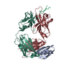
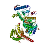
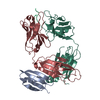
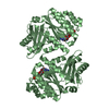
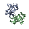
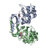
| Title | Crystal structure of SdgB (UDP and SD peptide-binding form) | ||||||||||||
 Components Components |
| ||||||||||||
 Keywords Keywords | TRANSFERASE / Glycosylation | ||||||||||||
| Function / homology |  Function and homology information Function and homology information | ||||||||||||
| Biological species |  Staphylococcus aureus subsp. aureus USA300 (bacteria) Staphylococcus aureus subsp. aureus USA300 (bacteria)synthetic construct (others) | ||||||||||||
| Method |  X-RAY DIFFRACTION / SYNCHROTRON / MOLECULAR REPLACEMENT / Resolution: 2.28 Å X-RAY DIFFRACTION / SYNCHROTRON / MOLECULAR REPLACEMENT / Resolution: 2.28 Å | ||||||||||||
 Authors Authors | Kim, D.-G. / Baek, I. / Lee, Y. / Kim, H.S. | ||||||||||||
| Funding support |  Korea, Republic Of, 3items Korea, Republic Of, 3items
| ||||||||||||
 Citation Citation | Journal: Acta Crystallogr D Struct Biol / Year: 2021 Title: Structural basis for SdgB- and SdgA-mediated glycosylation of staphylococcal adhesive proteins. Authors: Kim, D.G. / Baek, I. / Lee, Y. / Kim, H. / Kim, J.Y. / Bang, G. / Kim, S. / Yoon, H.J. / Han, B.W. / Suh, S.W. / Kim, H.S. | ||||||||||||
| History |
|
- Structure visualization
| Structure viewer | Molecule: MolmilJmol/JSmol |
|---|
- Downloads & links
-Download
| PDBx/mmCIF format | 7vfm.cif.gz | 215.1 KB | Display | PDBx/mmCIF format |
|---|---|---|---|---|
| PDB format | pdb7vfm.ent.gz | 169.9 KB | Display | PDB format |
| PDBx/mmJSON format | 7vfm.json.gz | Tree view | PDBx/mmJSON format | |
| Others |  Other downloads Other downloads |
-Validation report
| Arichive directory | https://data.pdbj.org/pub/pdb/validation_reports/vf/7vfmftp://data.pdbj.org/pub/pdb/validation_reports/vf/7vfm | HTTPS FTP |
|---|
-Related structure data
| Related structure data | 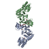 7ec1SC  7ec3C 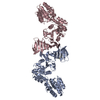 7ec6C  7ec7C  7vfkC  7vflC  7vfnC  7vfoC S: Starting model for refinement C: citing same article ( |
|---|---|
| Similar structure data |


-Links
 PDBj
PDBj


- Assembly
| Deposited unit | 
| ||||||||
|---|---|---|---|---|---|---|---|---|---|
| 1 | 
| ||||||||
| 2 | 
| ||||||||
| Unit cell |
|
-Components
| #1: Protein | Mass: 59647.316 Da / Num. of mol.: 2 Source method: isolated from a genetically manipulated source Source: (gene. exp.) Staphylococcus aureus subsp. aureus USA300 (bacteria)Strain: USA300 / Gene: SAUSA300_0550 / Production host: #2: Protein/peptide | Mass: 537.434 Da / Num. of mol.: 2 / Source method: obtained synthetically / Source: (synth.) synthetic construct (others) #3: Chemical |   Type: RNA linking / Mass: 404.161 Da / Num. of mol.: 2 / Source method: obtained synthetically / Formula: C9H14N2O12P2 / Feature type: SUBJECT OF INVESTIGATION / Comment: UDP*YM Type: RNA linking / Mass: 404.161 Da / Num. of mol.: 2 / Source method: obtained synthetically / Formula: C9H14N2O12P2 / Feature type: SUBJECT OF INVESTIGATION / Comment: UDP*YM#4: Water | ChemComp-HOH / |  Mass: 18.015 Da / Num. of mol.: 17 / Source method: isolated from a natural source / Formula: H2O Mass: 18.015 Da / Num. of mol.: 17 / Source method: isolated from a natural source / Formula: H2OHas ligand of interest | Y | |
|---|
-Experimental details
-Experiment
| Experiment | Method: X-RAY DIFFRACTION / Number of used crystals: 1 |
|---|
- Sample preparation
| Crystal | Density Matthews: 2.33 Å3/Da / Density % sol: 47.31 % |
|---|---|
| Crystal grow | Temperature: 287 K / Method: vapor diffusion, sitting drop Details: 0.2 M MgCl2, 0.1 M Tris-HCl (pH 8.5), and 25% (w/v) polyethylene glycol (PEG) 3,350 quick-soaked with 6.14 mM UDP and 2.45 mM 5-mer SD-repeat peptide (DSDSD) |
-Data collection
| Diffraction | Mean temperature: 100 K / Serial crystal experiment: N |
|---|---|
| Diffraction source | Source: SYNCHROTRON / Site: PAL/PLS / Beamline: 7A (6B, 6C1) / Wavelength: 0.9794 Å |
| Detector | Type: ADSC QUANTUM 270 / Detector: CCD / Date: Sep 29, 2014 |
| Radiation | Protocol: SINGLE WAVELENGTH / Monochromatic (M) / Laue (L): M / Scattering type: x-ray |
| Radiation wavelength | Wavelength: 0.9794 Å / Relative weight: 1 |
| Reflection | Resolution: 2.28→50 Å / Num. obs: 49800 / % possible obs: 99.6 % / Redundancy: 4.6 % / CC1/2: 0.995 / Net I/σ(I): 27.5 |
| Reflection shell | Resolution: 2.28→2.32 Å / Num. unique obs: 2418 / CC1/2: 0.71 |
- Processing
| Software |
| ||||||||||||||||||||||||||||||||||||||||||||||||||||||||||||||||||||||||||||||||||||||||||||||||||||||||||||||||||||||||||||||||||||||||||||
|---|---|---|---|---|---|---|---|---|---|---|---|---|---|---|---|---|---|---|---|---|---|---|---|---|---|---|---|---|---|---|---|---|---|---|---|---|---|---|---|---|---|---|---|---|---|---|---|---|---|---|---|---|---|---|---|---|---|---|---|---|---|---|---|---|---|---|---|---|---|---|---|---|---|---|---|---|---|---|---|---|---|---|---|---|---|---|---|---|---|---|---|---|---|---|---|---|---|---|---|---|---|---|---|---|---|---|---|---|---|---|---|---|---|---|---|---|---|---|---|---|---|---|---|---|---|---|---|---|---|---|---|---|---|---|---|---|---|---|---|---|---|
| Refinement | Method to determine structure: MOLECULAR REPLACEMENT Starting model: 7EC1 Resolution: 2.28→29.85 Å / SU ML: 0.39 / Cross valid method: THROUGHOUT / σ(F): 1.37 / Phase error: 32 / Stereochemistry target values: ML
| ||||||||||||||||||||||||||||||||||||||||||||||||||||||||||||||||||||||||||||||||||||||||||||||||||||||||||||||||||||||||||||||||||||||||||||
| Solvent computation | Shrinkage radii: 0.9 Å / VDW probe radii: 1.11 Å / Solvent model: FLAT BULK SOLVENT MODEL | ||||||||||||||||||||||||||||||||||||||||||||||||||||||||||||||||||||||||||||||||||||||||||||||||||||||||||||||||||||||||||||||||||||||||||||
| Displacement parameters | Biso max: 140.78 Å2 / Biso mean: 50.3535 Å2 / Biso min: 18.85 Å2 | ||||||||||||||||||||||||||||||||||||||||||||||||||||||||||||||||||||||||||||||||||||||||||||||||||||||||||||||||||||||||||||||||||||||||||||
| Refinement step | Cycle: final / Resolution: 2.28→29.85 Å
| ||||||||||||||||||||||||||||||||||||||||||||||||||||||||||||||||||||||||||||||||||||||||||||||||||||||||||||||||||||||||||||||||||||||||||||
| LS refinement shell | Refine-ID: X-RAY DIFFRACTION / Rfactor Rfree error: 0 / Total num. of bins used: 19
|
