TRANSFERASE / SALT STRESS / SODIUM TRANSPORT / ION HOMEOSTASIS
Function / homology
Function and homology information
plant-type vacuole membrane / response to salt stress / protein kinase activity / non-specific serine/threonine protein kinase / protein serine kinase activity / protein serine/threonine kinase activity / signal transduction / ATP binding / nucleus / cytoplasm Similarity search - Function
NAF domain / NAF/FISL domain / NAF domain / NAF domain profile. / Phosphorylase Kinase; domain 1 / Phosphorylase Kinase; domain 1 / Transferase(Phosphotransferase) domain 1 / Transferase(Phosphotransferase); domain 1 / Serine/threonine-protein kinase, active site / Serine/Threonine protein kinases active-site signature. ...NAF domain / NAF/FISL domain / NAF domain / NAF domain profile. / Phosphorylase Kinase; domain 1 / Phosphorylase Kinase; domain 1 / Transferase(Phosphotransferase) domain 1 / Transferase(Phosphotransferase); domain 1 / Serine/threonine-protein kinase, active site / Serine/Threonine protein kinases active-site signature. / Protein kinase domain / Serine/Threonine protein kinases, catalytic domain / Protein kinase, ATP binding site / Protein kinases ATP-binding region signature. / Protein kinase domain profile. / Protein kinase domain / Protein kinase-like domain superfamily / 2-Layer Sandwich / Orthogonal Bundle / Mainly Alpha / Alpha Beta Similarity search - Domain/homology
Resolution: 3.3→70.23 Å / Cor.coef. Fo:Fc: 0.891 / Cor.coef. Fo:Fc free: 0.875 / SU B: 42.659 / SU ML: 0.665 / Cross valid method: THROUGHOUT / ESU R Free: 0.747 / Stereochemistry target values: MAXIMUM LIKELIHOOD Details: HYDROGENS HAVE BEEN ADDED IN THE RIDING POSITIONS. U VALUES ARE REFINED INDIVIDUALLY
Rfactor
Num. reflection
% reflection
Selection details
Rfree
0.28312
810
5 %
RANDOM
Rwork
0.2706
-
-
-
obs
0.2712
15242
90.8 %
-
Solvent computation
Ion probe radii: 0.8 Å / Shrinkage radii: 0.8 Å / VDW probe radii: 1.2 Å / Solvent model: MASK
 Movie
Movie Controller


 Open data
 Basic information
 Components
 Keywords
 Function and homology information
 X-RAY DIFFRACTION /
  Authors
 Citation
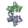
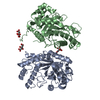
 Structure visualization
 Downloads & links
 Other downloads


 PDBj
PDBj Assembly
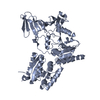
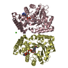
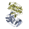
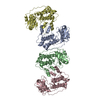


 Sample preparation
 / Beamline: P13 (MX1) / Wavelength: 0.9786
  Processing