 Movie
Movie Controller

[English] 日本語
 Yorodumi
- PDB-2i6t: Orthorhombic Structure of the LDH domain of Human Ubiquitin-conju... -
+ Open data

- Basic information
| Entry | Database: PDB / ID: 2i6t | ||||||
|---|---|---|---|---|---|---|---|
| Title | Orthorhombic Structure of the LDH domain of Human Ubiquitin-conjugating Enzyme E2-like Isoform A | ||||||
 Components Components | UBIQUITIN-CONJUGATING ENZYME E2-LIKE ISOFORM A | ||||||
 Keywords Keywords | UNKNOWN FUNCTION / L-lactate dehydrogenase / oxidoreductase / ubiquitin-protein ligase | ||||||
| Function / homology |  Function and homology information Function and homology informationESCRT I complex / carboxylic acid metabolic process / oxidoreductase activity, acting on the CH-OH group of donors, NAD or NADP as acceptor / endosome to lysosome transport / ubiquitin binding / protein transport / regulation of DNA-templated transcription / extracellular exosome / nucleus Similarity search - Function | ||||||
| Biological species |  Homo sapiens (human) Homo sapiens (human) | ||||||
| Method |  X-RAY DIFFRACTION / MOLECULAR REPLACEMENT / Resolution: 2.1 Å X-RAY DIFFRACTION / MOLECULAR REPLACEMENT / Resolution: 2.1 Å | ||||||
 Authors Authors | Walker, J.R. / Avvakumov, G.V. / Xue, S. / Newman, E.M. / Finerty Jr., P.J. / Butler-Cole, C. / Tempel, W. / Weigelt, J. / Sundstrom, M. / Arrowsmith, C.H. ...Walker, J.R. / Avvakumov, G.V. / Xue, S. / Newman, E.M. / Finerty Jr., P.J. / Butler-Cole, C. / Tempel, W. / Weigelt, J. / Sundstrom, M. / Arrowsmith, C.H. / Edwards, A.M. / Bochkarev, A. / Dhe-Paganon, S. | ||||||
 Citation Citation | Journal: To be Published Title: Structural Investigation into the L-lactate Dehydrogenase Domain of Human Ubiquitin-conjugating Enzyme E2-like Isoform A Authors: Avvakumov, G.V. / Walker, J.R. / Xue, S. / Newman, E.M. / Finerty Jr., P.J. / Butler-Cole, C. / Tempel, W. / Weigelt, J. / Sundstrom, M. / Arrowsmith, C.H. / Edwards, A.M. / Bochkarev, A. / Dhe-Paganon, S. #1: Journal: BIOCHIM.BIOPHYS.ACTA / Year: 2002 Title: Identification and characterization of UEV3, a human cDNA with similarities to inactive E2 ubiquitin-conjugating enzymes Authors: Kloor, M. / Bork, P. / Duwe, A. / Klaes, R. / von Knebel Doeberitz, M. / Ridder, R. | ||||||
| History |
|
- Structure visualization
| Structure viewer | Molecule: MolmilJmol/JSmol |
|---|
- Downloads & links
-Download
| PDBx/mmCIF format | 2i6t.cif.gz | 127.5 KB | Display | PDBx/mmCIF format |
|---|---|---|---|---|
| PDB format | pdb2i6t.ent.gz | 99.4 KB | Display | PDB format |
| PDBx/mmJSON format | 2i6t.json.gz | Tree view | PDBx/mmJSON format | |
| Others |  Other downloads Other downloads |
-Validation report
| Arichive directory | https://data.pdbj.org/pub/pdb/validation_reports/i6/2i6tftp://data.pdbj.org/pub/pdb/validation_reports/i6/2i6t | HTTPS FTP |
|---|
-Related structure data
| Related structure data |  1i10S S: Starting model for refinement |
|---|---|
| Similar structure data |


-Links
 PDBj
PDBj


- Assembly
| Deposited unit | 
| |||||||||
|---|---|---|---|---|---|---|---|---|---|---|
| 1 | 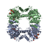
| |||||||||
| 2 |
| |||||||||
| Unit cell |
| |||||||||
| Components on special symmetry positions |
| |||||||||
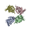
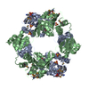
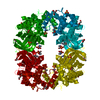
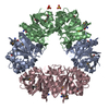
| Details | The biological assembly is a tetramer generated by the two fold axis: -x, -y, z. |
-Components
| #1: Protein | Mass: 32821.367 Da / Num. of mol.: 2 / Fragment: L-lactate Dehydrogenase Domain, residues 171-471 Source method: isolated from a genetically manipulated source Source: (gene. exp.) Homo sapiens (human) / Gene: UEVLD / Plasmid: pET28-LIC / Species (production host): Escherichia coli / Production host:  #2: Chemical | ChemComp-SO4 /   Mass: 96.063 Da / Num. of mol.: 4 / Source method: obtained synthetically / Formula: SO4 Mass: 96.063 Da / Num. of mol.: 4 / Source method: obtained synthetically / Formula: SO4#3: Chemical | ChemComp-GOL /   Mass: 92.094 Da / Num. of mol.: 7 / Source method: obtained synthetically / Formula: C3H8O3 Mass: 92.094 Da / Num. of mol.: 7 / Source method: obtained synthetically / Formula: C3H8O3#4: Water | ChemComp-HOH / |  Mass: 18.015 Da / Num. of mol.: 277 / Source method: isolated from a natural source / Formula: H2O Mass: 18.015 Da / Num. of mol.: 277 / Source method: isolated from a natural source / Formula: H2O |
|---|
-Experimental details
-Experiment
| Experiment | Method: X-RAY DIFFRACTION / Number of used crystals: 1 |
|---|
- Sample preparation
| Crystal | Density Matthews: 2.53 Å3/Da / Density % sol: 51.47 % |
|---|---|
| Crystal grow | Temperature: 298 K / Method: vapor diffusion, hanging drop Details: 26% PEG3350, 0.2 M ammonium sulfate, 0.1 M sodium cacodylate, pH 5.2 in protein buffer comprising of 20 mM Tris-HCl, pH 8.0, 500 mM NaCl, 5% glycerol, 10 mM dithiothreitol, VAPOR DIFFUSION, ...Details: 26% PEG3350, 0.2 M ammonium sulfate, 0.1 M sodium cacodylate, pH 5.2 in protein buffer comprising of 20 mM Tris-HCl, pH 8.0, 500 mM NaCl, 5% glycerol, 10 mM dithiothreitol, VAPOR DIFFUSION, HANGING DROP, temperature 298.0K |
-Data collection
| Diffraction | Mean temperature: 100 K |
|---|---|
| Diffraction source | Source: ROTATING ANODE / Type: RIGAKU / Wavelength: 1.54178 Å |
| Detector | Type: RIGAKU RAXIS IV++ / Detector: IMAGE PLATE / Date: Jul 19, 2006 |
| Radiation | Monochromator: GRAPHITE / Protocol: SINGLE WAVELENGTH / Monochromatic (M) / Laue (L): M / Scattering type: x-ray |
| Radiation wavelength | Wavelength: 1.54178 Å / Relative weight: 1 |
| Reflection | Resolution: 2.1→26.55 Å / Num. all: 39716 / Num. obs: 39716 / % possible obs: 99.9 % / Observed criterion σ(F): 0 / Observed criterion σ(I): -3 / Redundancy: 7 % / Rsym value: 0.18 / Net I/σ(I): 12.6 |
| Reflection shell | Resolution: 2.1→2.18 Å / Redundancy: 6.6 % / Mean I/σ(I) obs: 2.88 / Num. unique all: 3896 / Rsym value: 0.72 / % possible all: 100 |
- Processing
| Software |
| ||||||||||||||||||||||||||||||||||||||||||||||||||||||||||||||||||||||||||||||||||||||||||
|---|---|---|---|---|---|---|---|---|---|---|---|---|---|---|---|---|---|---|---|---|---|---|---|---|---|---|---|---|---|---|---|---|---|---|---|---|---|---|---|---|---|---|---|---|---|---|---|---|---|---|---|---|---|---|---|---|---|---|---|---|---|---|---|---|---|---|---|---|---|---|---|---|---|---|---|---|---|---|---|---|---|---|---|---|---|---|---|---|---|---|---|
| Refinement | Method to determine structure: MOLECULAR REPLACEMENT Starting model: PDB ENTRY 1I10 Resolution: 2.1→26.55 Å / Cor.coef. Fo:Fc: 0.954 / Cor.coef. Fo:Fc free: 0.934 / SU B: 4.134 / SU ML: 0.112 / Cross valid method: THROUGHOUT / ESU R: 0.192 / ESU R Free: 0.174 / Stereochemistry target values: MAXIMUM LIKELIHOOD / Details: HYDROGENS HAVE BEEN ADDED IN THE RIDING POSITIONS
| ||||||||||||||||||||||||||||||||||||||||||||||||||||||||||||||||||||||||||||||||||||||||||
| Solvent computation | Ion probe radii: 0.8 Å / Shrinkage radii: 0.8 Å / VDW probe radii: 1.4 Å / Solvent model: BABINET MODEL WITH MASK | ||||||||||||||||||||||||||||||||||||||||||||||||||||||||||||||||||||||||||||||||||||||||||
| Displacement parameters | Biso mean: 23.562 Å2
| ||||||||||||||||||||||||||||||||||||||||||||||||||||||||||||||||||||||||||||||||||||||||||
| Refinement step | Cycle: LAST / Resolution: 2.1→26.55 Å
| ||||||||||||||||||||||||||||||||||||||||||||||||||||||||||||||||||||||||||||||||||||||||||
| Refine LS restraints |
| ||||||||||||||||||||||||||||||||||||||||||||||||||||||||||||||||||||||||||||||||||||||||||
| LS refinement shell | Resolution: 2.1→2.154 Å / Total num. of bins used: 20
|
