Mass: 18.015 Da / Num. of mol.: 37 / Source method: isolated from a natural source / Formula: H2O
Compound details
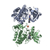
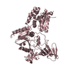
ACCORDING TO THE AUTHORS CHAIN C LIKELY REPRESENTS PART OF THE PROTEIN CONSTRUCT'S N-TERMINUS. ...ACCORDING TO THE AUTHORS CHAIN C LIKELY REPRESENTS PART OF THE PROTEIN CONSTRUCT'S N-TERMINUS. DENSITY WAS DISCONTINUOUS AND THE AMINO ACID SEQUENCE OF THE FRAGMENT COULD NOT BE ASSIGNED.
-
Experimental details
-
Experiment
Experiment
Method: X-RAY DIFFRACTION / Number of used crystals: 1
-
Sample preparation
Crystal
Density Matthews: 2.39 Å3/Da / Density % sol: 48.7 %
Crystal grow
Temperature: 291 K / Method: vapor diffusion, hanging drop / pH: 8 Details: 16% peg-2000 mme, 0.1M TRIS, 0.2M TMAO, pH 8, vapor diffusion, hanging drop, temperature 291K
Protocol: SINGLE WAVELENGTH / Monochromatic (M) / Laue (L): M / Scattering type: x-ray
Radiation wavelength
Wavelength: 0.9999 Å / Relative weight: 1
Reflection
Resolution: 2.4→30 Å / Num. obs: 27657 / % possible obs: 99.9 % / Redundancy: 9.8 % / Rmerge(I) obs: 0.045 / Χ2: 1.038 / Net I/σ(I): 15
Reflection shell
Resolution (Å)
Redundancy (%)
Rmerge(I) obs
Num. unique all
Χ2
Diffraction-ID
% possible all
2.4-2.49
7.6
0.577
2703
0.965
1
99.9
2.49-2.59
8.9
0.408
2686
0.962
1
100
2.59-2.7
9.5
0.296
2723
0.982
1
100
2.7-2.85
10
0.191
2737
0.992
1
100
2.85-3.02
10.3
0.127
2735
0.97
1
100
3.02-3.26
10.5
0.083
2739
0.982
1
100
3.26-3.58
10.6
0.048
2771
0.983
1
100
3.58-4.1
10.6
0.038
2787
1.271
1
100
4.1-5.16
10.3
0.033
2814
1.442
1
100
5.16-30
9.7
0.019
2962
0.777
1
99.2
-
Phasing
Phasing
Method: sad
-
Processing
Software
Name
Version
Classification
NB
DENZO
datareduction
SCALEPACK
datascaling
SOLVE
phasing
RESOLVE
phasing
REFMAC
5.2.0019
refinement
PDB_EXTRACT
3.005
dataextraction
HKL-2000
datascaling
Refinement
Method to determine structure: SAD Starting model: For phasing, native crystals were incubated over-night in an artificial mother liquor containing 0.001 M thimerosal. Resolution: 2.402→28.083 Å / Cor.coef. Fo:Fc: 0.94 / Cor.coef. Fo:Fc free: 0.928 / WRfactor Rfree: 0.259 / WRfactor Rwork: 0.224 / SU B: 19.659 / SU ML: 0.216 / TLS residual ADP flag: LIKELY RESIDUAL / Cross valid method: THROUGHOUT / σ(F): 0 / ESU R: 0.362 / ESU R Free: 0.255 / Stereochemistry target values: MAXIMUM LIKELIHOOD Details: HYDROGENS HAVE BEEN ADDED IN THE RIDING POSITIONS. Atomic B-factors are residuals from TLS refinement. Chain C likely represents part of the protein construct's N-terminus. Density was ...Details: HYDROGENS HAVE BEEN ADDED IN THE RIDING POSITIONS. Atomic B-factors are residuals from TLS refinement. Chain C likely represents part of the protein construct's N-terminus. Density was discontinuous and the amino acid sequence of the fragment could not be assigned. O, COOT, MOLPROBITY were also used for refinement.
Rfactor
Num. reflection
% reflection
Selection details
Rfree
0.262
1390
5.037 %
RANDOM
Rwork
0.227
-
-
-
obs
0.228
27598
99.779 %
-
Solvent computation
Ion probe radii: 0.8 Å / Shrinkage radii: 0.8 Å / VDW probe radii: 1.2 Å / Solvent model: MASK BULK SOLVENT
Displacement parameters
Biso mean: 47.297 Å2
Baniso -1
Baniso -2
Baniso -3
1-
0.953 Å2
0.476 Å2
0 Å2
2-
-
0.953 Å2
0 Å2
3-
-
-
-1.429 Å2
Refinement step
Cycle: LAST / Resolution: 2.402→28.083 Å
Protein
Nucleic acid
Ligand
Solvent
Total
Num. atoms
4078
0
19
37
4134
Refine LS restraints
Refine-ID
Type
Dev ideal
Dev ideal target
Number
X-RAY DIFFRACTION
r_bond_refined_d
0.013
0.022
4159
X-RAY DIFFRACTION
r_bond_other_d
0.001
0.02
2741
X-RAY DIFFRACTION
r_angle_refined_deg
1.213
1.97
5661
X-RAY DIFFRACTION
r_angle_other_deg
0.899
3
6697
X-RAY DIFFRACTION
r_dihedral_angle_1_deg
5.576
5
523
X-RAY DIFFRACTION
r_dihedral_angle_2_deg
34.655
23.728
169
X-RAY DIFFRACTION
r_dihedral_angle_3_deg
14.896
15
675
X-RAY DIFFRACTION
r_dihedral_angle_4_deg
17.721
15
25
X-RAY DIFFRACTION
r_chiral_restr
0.068
0.2
667
X-RAY DIFFRACTION
r_gen_planes_refined
0.004
0.02
4590
X-RAY DIFFRACTION
r_gen_planes_other
0.001
0.02
828
X-RAY DIFFRACTION
r_nbd_refined
0.196
0.2
945
X-RAY DIFFRACTION
r_nbd_other
0.177
0.2
2644
X-RAY DIFFRACTION
r_nbtor_refined
0.175
0.2
2037
X-RAY DIFFRACTION
r_nbtor_other
0.087
0.2
2059
X-RAY DIFFRACTION
r_xyhbond_nbd_refined
0.139
0.2
114
X-RAY DIFFRACTION
r_symmetry_vdw_refined
0.201
0.2
14
X-RAY DIFFRACTION
r_symmetry_vdw_other
0.274
0.2
56
X-RAY DIFFRACTION
r_symmetry_hbond_refined
0.088
0.2
3
X-RAY DIFFRACTION
r_mcbond_it
1.932
2
2713
X-RAY DIFFRACTION
r_mcbond_other
0.454
2
1056
X-RAY DIFFRACTION
r_mcangle_it
2.886
3
4239
X-RAY DIFFRACTION
r_scbond_it
1.923
2
1656
X-RAY DIFFRACTION
r_scangle_it
2.695
3
1422
LS refinement shell
Refine-ID: X-RAY DIFFRACTION / Total num. of bins used: 20
Resolution (Å)
Rfactor Rfree
Num. reflection Rfree
Rfactor Rwork
Num. reflection Rwork
Num. reflection all
% reflection obs (%)
2.402-2.464
0.402
110
0.285
1846
1992
98.193
2.464-2.531
0.381
94
0.27
1824
1919
99.948
2.531-2.604
0.306
86
0.265
1813
1900
99.947
2.604-2.683
0.314
97
0.26
1766
1863
100
2.683-2.771
0.356
93
0.252
1689
1782
100
2.771-2.867
0.287
88
0.241
1644
1733
99.942
2.867-2.974
0.354
85
0.246
1601
1686
100
2.974-3.094
0.33
79
0.247
1525
1604
100
3.094-3.23
0.246
73
0.253
1490
1563
100
3.23-3.385
0.286
75
0.232
1433
1508
100
3.385-3.565
0.238
74
0.217
1333
1407
100
3.565-3.778
0.254
60
0.222
1294
1354
100
3.778-4.033
0.269
63
0.208
1204
1267
100
4.033-4.349
0.23
60
0.192
1140
1200
100
4.349-4.753
0.218
53
0.187
1045
1098
100
4.753-5.295
0.215
48
0.208
977
1025
100
5.295-6.078
0.307
50
0.263
855
905
100
6.078-7.358
0.276
51
0.245
741
792
100
7.358-10.067
0.168
29
0.194
605
634
100
10.067-28.083
0.18
22
0.238
383
427
94.848
Refinement TLS params.
Method: refined / Origin x: -20.2896 Å / Origin y: 29.0521 Å / Origin z: -16.9473 Å
11
12
13
21
22
23
31
32
33
T
-0.0253 Å2
-0.1049 Å2
0.0063 Å2
-
-0.3137 Å2
0.0071 Å2
-
-
-0.2068 Å2
L
1.1305 °2
-0.0362 °2
-0.5348 °2
-
1.4759 °2
-0.3424 °2
-
-
2.6048 °2
S
-0.1482 Å °
0.0084 Å °
-0.0426 Å °
-0.2278 Å °
0.0892 Å °
-0.1182 Å °
0.4391 Å °
-0.0207 Å °
0.059 Å °
Refinement TLS group
ID
Refine-ID
Refine TLS-ID
Auth asym-ID
Auth seq-ID
1
X-RAY DIFFRACTION
1
X
1563 - 2129
2
X-RAY DIFFRACTION
1
C
1007 - 1033
+
About Yorodumi
-
News
-
Feb 9, 2022. New format data for meta-information of EMDB entries
New format data for meta-information of EMDB entries
Version 3 of the EMDB header file is now the official format.
The previous official version 1.9 will be removed from the archive.
In the structure databanks used in Yorodumi, some data are registered as the other names, "COVID-19 virus" and "2019-nCoV". Here are the details of the virus and the list of structure data.
Jan 31, 2019. EMDB accession codes are about to change! (news from PDBe EMDB page)
EMDB accession codes are about to change! (news from PDBe EMDB page)
The allocation of 4 digits for EMDB accession codes will soon come to an end. Whilst these codes will remain in use, new EMDB accession codes will include an additional digit and will expand incrementally as the available range of codes is exhausted. The current 4-digit format prefixed with “EMD-” (i.e. EMD-XXXX) will advance to a 5-digit format (i.e. EMD-XXXXX), and so on. It is currently estimated that the 4-digit codes will be depleted around Spring 2019, at which point the 5-digit format will come into force.
The EM Navigator/Yorodumi systems omit the EMD- prefix.
Related info.:Q: What is EMD? / ID/Accession-code notation in Yorodumi/EM Navigator
Yorodumi is a browser for structure data from EMDB, PDB, SASBDB, etc.
This page is also the successor to EM Navigator detail page, and also detail information page/front-end page for Omokage search.
The word "yorodu" (or yorozu) is an old Japanese word meaning "ten thousand". "mi" (miru) is to see.
Related info.:EMDB / PDB / SASBDB / Comparison of 3 databanks / Yorodumi Search / Aug 31, 2016. New EM Navigator & Yorodumi / Yorodumi Papers / Jmol/JSmol / Function and homology information / Changes in new EM Navigator and Yorodumi
 Movie
Movie Controller


 Open data
 Basic information
 Components
 Keywords
 Function and homology information
 Homo sapiens (human)
 X-RAY DIFFRACTION /
  Authors
 Citation
 Structure visualization
 Downloads & links
 Other downloads
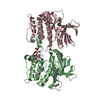

 PDBj
PDBj


 Assembly

 Spodoptera frugiperda (fall armyworm) / Strain (production host): SF9 / References: UniProt: O43157
 Num. of mol.: 19 / Source method: obtained synthetically
 Mass: 18.015 Da / Num. of mol.: 37 / Source method: isolated from a natural source / Formula: H2O
 Sample preparation
 / Beamline: X25 / Wavelength: 0.9999 Å
 Processing