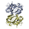
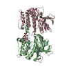
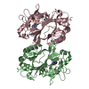
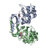
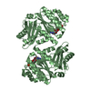
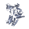
BIOMOLECULE: 1 THIS ENTRY CONTAINS THE CRYSTALLOGRAPHIC ASYMMETRIC UNIT WHICH CONSISTS OF 1 ... BIOMOLECULE: 1 THIS ENTRY CONTAINS THE CRYSTALLOGRAPHIC ASYMMETRIC UNIT WHICH CONSISTS OF 1 CHAIN(S). SEE REMARK 350 FOR INFORMATION ON GENERATING THE BIOLOGICAL MOLECULE(S). STATIC LIGHT SCATTERING AND ANALYTICAL SIZE EXCLUSION CHROMATOGRAPHY MEASUREMENTS INDICATE THAT THE PROTEIN IS A DIMER IN SOLUTION.
Resolution: 2.3→19.8 Å / Cor.coef. Fo:Fc: 0.945 / Cor.coef. Fo:Fc free: 0.906 / SU B: 14.526 / SU ML: 0.18 / TLS residual ADP flag: LIKELY RESIDUAL / Cross valid method: THROUGHOUT / σ(F): 0 / ESU R: 0.22 / ESU R Free: 0.21 / Stereochemistry target values: MAXIMUM LIKELIHOOD Details: 1. HYDROGENS HAVE BEEN ADDED IN THE RIDING POSITIONS. 2. ATOM RECORD CONTAINS RESIDUAL B FACTORS ONLY 3. A MET-INHIBITION PROTOCOL WAS USED FOR SELENOMETHIONINE INCORPORATION DURING PROTEIN ...Details: 1. HYDROGENS HAVE BEEN ADDED IN THE RIDING POSITIONS. 2. ATOM RECORD CONTAINS RESIDUAL B FACTORS ONLY 3. A MET-INHIBITION PROTOCOL WAS USED FOR SELENOMETHIONINE INCORPORATION DURING PROTEIN EXPRESSION. THE OCCUPANCY OF THE SE ATOMS IN THE MSE RESIDUES WAS REDUCED TO 0.70 FOR THE REDUCED SCATTERING POWER DUE TO PARTIAL S-MET INCORPORATION. 4. RESIDUES 1-5 AND 311 ARE DISORDERED AND NOT INCLUDED IN THE MODEL. 5. THE PROTEIN WAS CO- CRYSTALLIZED IN THE PRESENCE OF FE PROTOPORPHYRIN (IX).
Rfactor
Num. reflection
% reflection
Selection details
Rfree
0.266
1269
5.2 %
RANDOM
Rwork
0.205
-
-
-
obs
0.208
24207
99.88 %
-
Solvent computation
Ion probe radii: 0.8 Å / Shrinkage radii: 0.8 Å / VDW probe radii: 1.2 Å / Solvent model: MASK
Displacement parameters
Biso mean: 33.304 Å2
Baniso -1
Baniso -2
Baniso -3
1-
-1.39 Å2
0 Å2
0 Å2
2-
-
-1.39 Å2
0 Å2
3-
-
-
2.78 Å2
Refinement step
Cycle: LAST / Resolution: 2.3→19.8 Å
Protein
Nucleic acid
Ligand
Solvent
Total
Num. atoms
2441
0
56
112
2609
Refine LS restraints
Refine-ID
Type
Dev ideal
Dev ideal target
Number
X-RAY DIFFRACTION
r_bond_refined_d
0.013
0.022
2566
X-RAY DIFFRACTION
r_bond_other_d
0.002
0.02
2221
X-RAY DIFFRACTION
r_angle_refined_deg
1.357
2.001
3474
X-RAY DIFFRACTION
r_angle_other_deg
0.763
3
5160
X-RAY DIFFRACTION
r_dihedral_angle_1_deg
6.456
5
307
X-RAY DIFFRACTION
r_dihedral_angle_2_deg
36.049
24.254
134
X-RAY DIFFRACTION
r_dihedral_angle_3_deg
13.869
15
421
X-RAY DIFFRACTION
r_dihedral_angle_4_deg
16.274
15
16
X-RAY DIFFRACTION
r_chiral_restr
0.076
0.2
348
X-RAY DIFFRACTION
r_gen_planes_refined
0.001
0.02
2888
X-RAY DIFFRACTION
r_gen_planes_other
0
0.02
530
X-RAY DIFFRACTION
r_nbd_refined
0.203
0.2
567
X-RAY DIFFRACTION
r_nbd_other
0.19
0.2
2213
X-RAY DIFFRACTION
r_nbtor_refined
0.18
0.2
1217
X-RAY DIFFRACTION
r_nbtor_other
0.087
0.2
1358
X-RAY DIFFRACTION
r_xyhbond_nbd_refined
0.134
0.2
107
X-RAY DIFFRACTION
r_symmetry_vdw_refined
0.255
0.2
11
X-RAY DIFFRACTION
r_symmetry_vdw_other
0.24
0.2
46
X-RAY DIFFRACTION
r_symmetry_hbond_refined
0.292
0.2
9
X-RAY DIFFRACTION
r_mcbond_it
1.33
3
1525
X-RAY DIFFRACTION
r_mcbond_other
0.329
3
621
X-RAY DIFFRACTION
r_mcangle_it
2.281
5
2446
X-RAY DIFFRACTION
r_scbond_it
4.387
8
1075
X-RAY DIFFRACTION
r_scangle_it
5.78
11
1027
LS refinement shell
Resolution: 2.3→2.359 Å / Total num. of bins used: 20
Rfactor
Num. reflection
% reflection
Rfree
0.374
88
-
Rwork
0.292
1650
-
obs
-
1738
99.94 %
Refinement TLS params.
Method: refined / Origin x: -19.721 Å / Origin y: -19.058 Å / Origin z: -18.559 Å
11
12
13
21
22
23
31
32
33
T
0.0566 Å2
-0.0344 Å2
0.0412 Å2
-
-0.0962 Å2
0.0093 Å2
-
-
-0.1493 Å2
L
1.7409 °2
-0.6528 °2
0.0518 °2
-
3.598 °2
-0.5402 °2
-
-
2.0832 °2
S
0.0579 Å °
0.3242 Å °
0.047 Å °
-0.4916 Å °
-0.0705 Å °
-0.3376 Å °
0.113 Å °
0.2124 Å °
0.0126 Å °
Refinement TLS group
Selection: ALL
+
About Yorodumi
-
News
-
Feb 9, 2022. New format data for meta-information of EMDB entries
New format data for meta-information of EMDB entries
Version 3 of the EMDB header file is now the official format.
The previous official version 1.9 will be removed from the archive.
In the structure databanks used in Yorodumi, some data are registered as the other names, "COVID-19 virus" and "2019-nCoV". Here are the details of the virus and the list of structure data.
Jan 31, 2019. EMDB accession codes are about to change! (news from PDBe EMDB page)
EMDB accession codes are about to change! (news from PDBe EMDB page)
The allocation of 4 digits for EMDB accession codes will soon come to an end. Whilst these codes will remain in use, new EMDB accession codes will include an additional digit and will expand incrementally as the available range of codes is exhausted. The current 4-digit format prefixed with “EMD-” (i.e. EMD-XXXX) will advance to a 5-digit format (i.e. EMD-XXXXX), and so on. It is currently estimated that the 4-digit codes will be depleted around Spring 2019, at which point the 5-digit format will come into force.
The EM Navigator/Yorodumi systems omit the EMD- prefix.
Related info.:Q: What is EMD? / ID/Accession-code notation in Yorodumi/EM Navigator
Yorodumi is a browser for structure data from EMDB, PDB, SASBDB, etc.
This page is also the successor to EM Navigator detail page, and also detail information page/front-end page for Omokage search.
The word "yorodu" (or yorozu) is an old Japanese word meaning "ten thousand". "mi" (miru) is to see.
Related info.:EMDB / PDB / SASBDB / Comparison of 3 databanks / Yorodumi Search / Aug 31, 2016. New EM Navigator & Yorodumi / Yorodumi Papers / Jmol/JSmol / Function and homology information / Changes in new EM Navigator and Yorodumi
 Movie
Movie Controller

 Yorodumi
 Open data
 Basic information
 Components
 Keywords
 Function and homology information
 Shewanella oneidensis (bacteria)
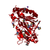
 X-RAY DIFFRACTION /
  Authors
 Citation
 Structure visualization
 Downloads & links
 Other downloads

 PDBj
PDBj
 Assembly
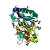


 Mass: 22.990 Da / Num. of mol.: 1 / Source method: obtained synthetically / Formula: Na
 Mass: 616.487 Da / Num. of mol.: 1 / Source method: obtained synthetically / Formula: C34H32FeN4O4
 Mass: 62.068 Da / Num. of mol.: 2 / Source method: obtained synthetically / Formula: C2H6O2
 Mass: 60.095 Da / Num. of mol.: 1 / Source method: obtained synthetically / Formula: C3H8O
 Sample preparation
 / Beamline: BL11-1
 Processing