 Movie
Movie Controller


+ Open data

- Basic information
| Entry | Database: PDB / ID: 7v8p | ||||||
|---|---|---|---|---|---|---|---|
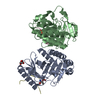
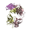
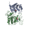
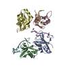
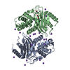
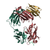
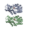
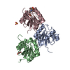
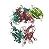
| Title | Crystal Structure of the MukE dimer | ||||||
 Components Components | Chromosome partition protein MukE | ||||||
 Keywords Keywords | NUCLEAR PROTEIN / Structural maintenance of chromosomes / Chromatin remodeling | ||||||
| Function / homology | Arc Repressor Mutant, subunit A - #2250 / Arc Repressor Mutant, subunit A / Orthogonal Bundle / Mainly Alpha / :  Function and homology information Function and homology information | ||||||
| Biological species |  Shigella flexneri (bacteria) Shigella flexneri (bacteria) | ||||||
| Method |  X-RAY DIFFRACTION / SYNCHROTRON / MOLECULAR REPLACEMENT / Resolution: 2.44 Å X-RAY DIFFRACTION / SYNCHROTRON / MOLECULAR REPLACEMENT / Resolution: 2.44 Å | ||||||
 Authors Authors | Qian, J.W. / Guo, L. | ||||||
| Funding support |  China, 1items China, 1items
| ||||||
 Citation Citation | Journal: Biochem.Biophys.Res.Commun. / Year: 2021 Title: Crystal structure of the chromosome partition protein MukE homodimer. Authors: Qian, J.W. / Wang, X.Y. / Deng, K. / Li, D.F. / Guo, L. | ||||||
| History |
|
- Structure visualization
| Structure viewer | Molecule: MolmilJmol/JSmol |
|---|
- Downloads & links
-Download
| PDBx/mmCIF format | 7v8p.cif.gz | 96.1 KB | Display | PDBx/mmCIF format |
|---|---|---|---|---|
| PDB format | pdb7v8p.ent.gz | 72 KB | Display | PDB format |
| PDBx/mmJSON format | 7v8p.json.gz | Tree view | PDBx/mmJSON format | |
| Others |  Other downloads Other downloads |
-Validation report
| Summary document | 7v8p_validation.pdf.gz | 425.9 KB | Display | wwPDB validaton report |
|---|---|---|---|---|
| Full document | 7v8p_full_validation.pdf.gz | 427.5 KB | Display | |
| Data in XML | 7v8p_validation.xml.gz | 8.9 KB | Display | |
| Data in CIF | 7v8p_validation.cif.gz | 10.8 KB | Display | |
| Arichive directory | https://data.pdbj.org/pub/pdb/validation_reports/v8/7v8pftp://data.pdbj.org/pub/pdb/validation_reports/v8/7v8p | HTTPS FTP |
-Related structure data
| Related structure data |  3euhS S: Starting model for refinement |
|---|---|
| Similar structure data |

-Links
 PDBj
PDBj- Assembly
| Deposited unit | 
| ||||||||
|---|---|---|---|---|---|---|---|---|---|
| 1 | 
| ||||||||
| Unit cell |
|
-Components
| #1: Protein | Mass: 25989.363 Da / Num. of mol.: 1 Source method: isolated from a genetically manipulated source Source: (gene. exp.) Shigella flexneri (bacteria)Gene: mukE, CQA91_05990, DQP17_14640, NCTC8524_01712, NCTC9783_04161, SAMEA3710514_03893, SAMEA3710568_03348 Production host: References: UniProt: A0A383JZS2 |
|---|---|
| #2: Water | ChemComp-HOH /  Mass: 18.015 Da / Num. of mol.: 4 / Source method: isolated from a natural source / Formula: H2O Mass: 18.015 Da / Num. of mol.: 4 / Source method: isolated from a natural source / Formula: H2O |
-Experimental details
-Experiment
| Experiment | Method: X-RAY DIFFRACTION / Number of used crystals: 1 |
|---|
- Sample preparation
| Crystal | Density Matthews: 2.26 Å3/Da / Density % sol: 45.53 % |
|---|---|
| Crystal grow | Temperature: 298 K / Method: vapor diffusion Details: 0.2 M Sodium chloride;0.1 M IBS pH 8.5;25% w/v Polyethylene glycol 3,350 |
-Data collection
| Diffraction | Mean temperature: 95 K / Serial crystal experiment: N | ||||||||||||||||||||||||||||||
|---|---|---|---|---|---|---|---|---|---|---|---|---|---|---|---|---|---|---|---|---|---|---|---|---|---|---|---|---|---|---|---|
| Diffraction source | Source: SYNCHROTRON / Site: Photon Factory  / Beamline: BL-6A / Wavelength: 0.9792 Å / Beamline: BL-6A / Wavelength: 0.9792 Å | ||||||||||||||||||||||||||||||
| Detector | Type: ADSC QUANTUM 4 / Detector: CCD / Date: Aug 11, 2007 | ||||||||||||||||||||||||||||||
| Radiation | Protocol: SINGLE WAVELENGTH / Monochromatic (M) / Laue (L): M / Scattering type: x-ray | ||||||||||||||||||||||||||||||
| Radiation wavelength | Wavelength: 0.9792 Å / Relative weight: 1 | ||||||||||||||||||||||||||||||
| Reflection | Resolution: 2.439→55.64 Å / Num. obs: 8635 / % possible obs: 99.3 % / Redundancy: 7.4 % / CC1/2: 0.998 / Rmerge(I) obs: 0.094 / Rpim(I) all: 0.037 / Rrim(I) all: 0.101 / Net I/σ(I): 12 | ||||||||||||||||||||||||||||||
| Reflection shell | Diffraction-ID: 1
|
- Processing
| Software |
| ||||||||||||||||||||||||||||||||||||||||
|---|---|---|---|---|---|---|---|---|---|---|---|---|---|---|---|---|---|---|---|---|---|---|---|---|---|---|---|---|---|---|---|---|---|---|---|---|---|---|---|---|---|
| Refinement | Method to determine structure: MOLECULAR REPLACEMENT Starting model: 3EUH Resolution: 2.44→32.656 Å / SU ML: 0.17 / Cross valid method: THROUGHOUT / σ(F): 1.35 / Phase error: 33.61 / Stereochemistry target values: ML
| ||||||||||||||||||||||||||||||||||||||||
| Solvent computation | Shrinkage radii: 0.9 Å / VDW probe radii: 1.11 Å / Solvent model: FLAT BULK SOLVENT MODEL | ||||||||||||||||||||||||||||||||||||||||
| Displacement parameters | Biso max: 133.8 Å2 / Biso mean: 68.365 Å2 / Biso min: 30.14 Å2 | ||||||||||||||||||||||||||||||||||||||||
| Refinement step | Cycle: final / Resolution: 2.44→32.656 Å
| ||||||||||||||||||||||||||||||||||||||||
| LS refinement shell | Refine-ID: X-RAY DIFFRACTION / Rfactor Rfree error: 0
| ||||||||||||||||||||||||||||||||||||||||
| Refinement TLS params. | Method: refined / Origin x: -0.8302 Å / Origin y: 34.4444 Å / Origin z: -16.9704 Å
| ||||||||||||||||||||||||||||||||||||||||
| Refinement TLS group | Selection details: (chain 'A' and resid 1 through 220) |
