Redundancy: 4.9 % / Av σ(I) over netI: 31.82 / Number: 196180 / Rmerge(I) obs: 0.09 / Χ2: 1.67 / D res high: 3.1 Å / D res low: 30 Å / Num. obs: 39887 / % possible obs: 97.3
Diffraction reflection shell
Highest resolution (Å)
Lowest resolution (Å)
% possible obs (%)
ID
Rmerge(I) obs
Chi squared
Redundancy
6.66
30
99.4
1
0.058
2.488
5.9
5.29
6.66
98.7
1
0.09
2.074
5.5
4.63
5.29
99
1
0.09
2.052
5.6
4.21
4.63
98.6
1
0.094
1.88
5.5
3.9
4.21
97.6
1
0.107
1.688
5.3
3.67
3.9
97.2
1
0.129
1.434
4.8
3.49
3.67
96.7
1
0.146
1.25
4.6
3.34
3.49
95.9
1
0.169
1.084
4.2
3.21
3.34
95.5
1
0.188
0.78
3.9
3.1
3.21
93.7
1
0.218
0.676
3.6
Reflection
Resolution: 2.7→30 Å / Num. obs: 59926 / % possible obs: 97.1 % / Redundancy: 6.1 % / Rmerge(I) obs: 0.065 / Χ2: 1.675 / Net I/σ(I): 31.821
Reflection shell
Resolution (Å)
Redundancy (%)
Rmerge(I) obs
Num. unique all
Χ2
% possible all
2.7-2.8
3.8
0.227
5609
0.495
92.6
2.8-2.91
4.3
0.198
5760
0.55
94.8
2.91-3.04
4.9
0.16
5855
0.69
95.9
3.04-3.2
5.4
0.132
5921
0.882
97.1
3.2-3.4
6.1
0.108
6014
1.192
98.1
3.4-3.66
6.4
0.092
5968
1.622
97.5
3.66-4.03
7
0.079
6026
1.965
97.9
4.03-4.61
7.6
0.066
6174
2.306
99.2
4.61-5.8
7.7
0.059
6200
2.375
99.5
5.8-30
7.3
0.048
6399
2.502
98.3
-
Processing
Software
Name
Version
Classification
NB
DENZO
datareduction
SCALEPACK
datascaling
CNS
refinement
PDB_EXTRACT
3.006
dataextraction
MLPHARE
phasing
Refinement
Method to determine structure: SIRAS / Resolution: 2.9→20 Å / Occupancy max: 1 / Occupancy min: 1 / FOM work R set: 0.834 / σ(F): 0
In the structure databanks used in Yorodumi, some data are registered as the other names, "COVID-19 virus" and "2019-nCoV". Here are the details of the virus and the list of structure data.
Jan 31, 2019. EMDB accession codes are about to change! (news from PDBe EMDB page)
EMDB accession codes are about to change! (news from PDBe EMDB page)
The allocation of 4 digits for EMDB accession codes will soon come to an end. Whilst these codes will remain in use, new EMDB accession codes will include an additional digit and will expand incrementally as the available range of codes is exhausted. The current 4-digit format prefixed with “EMD-” (i.e. EMD-XXXX) will advance to a 5-digit format (i.e. EMD-XXXXX), and so on. It is currently estimated that the 4-digit codes will be depleted around Spring 2019, at which point the 5-digit format will come into force.
The EM Navigator/Yorodumi systems omit the EMD- prefix.
Related info.:Q: What is EMD? / ID/Accession-code notation in Yorodumi/EM Navigator
Yorodumi is a browser for structure data from EMDB, PDB, SASBDB, etc.
This page is also the successor to EM Navigator detail page, and also detail information page/front-end page for Omokage search.
The word "yorodu" (or yorozu) is an old Japanese word meaning "ten thousand". "mi" (miru) is to see.
Related info.:EMDB / PDB / SASBDB / Comparison of 3 databanks / Yorodumi Search / Aug 31, 2016. New EM Navigator & Yorodumi / Yorodumi Papers / Jmol/JSmol / Function and homology information / Changes in new EM Navigator and Yorodumi
 Movie
Movie Controller


 Open data
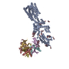
 Basic information
 Components
 Keywords
 Function and homology information
 X-RAY DIFFRACTION /
  Authors
 Citation
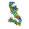
 Structure visualization
 Downloads & links
 Other downloads
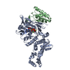


 PDBj
PDBj

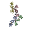
 Assembly
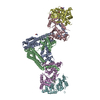

 Type: peptide linking / Mass: 75.067 Da / Num. of mol.: 4 / Source method: obtained synthetically / Formula: C2H5NO2
 Mass: 18.015 Da / Num. of mol.: 57 / Source method: isolated from a natural source / Formula: H2O
 Sample preparation


 Processing