 Movie
Movie Controller

[English] 日本語
 Yorodumi
- PDB-2xnk: Structure and function of the Rad9-binding region of the DNA dama... -
+ Open data

- Basic information
| Entry | Database: PDB / ID: 2xnk | ||||||
|---|---|---|---|---|---|---|---|
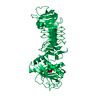
| Title | Structure and function of the Rad9-binding region of the DNA damage checkpoint adaptor TopBP1 | ||||||
 Components Components | DNA TOPOISOMERASE 2-BINDING PROTEIN 1 | ||||||
 Keywords Keywords | ISOMERASE / PHOSPHORYLATION / PROTEIN-PROTEIN INTERACTION / DNA REPAIR | ||||||
| Function / homology |  Function and homology information Function and homology informationbroken chromosome clustering / BRCA1-B complex / phosphorylation-dependent protein binding / homologous recombination / DNA replication checkpoint signaling / double-strand break repair via classical nonhomologous end joining / double-strand break repair via alternative nonhomologous end joining / mitotic DNA replication checkpoint signaling / protein localization to site of double-strand break / chromatin-protein adaptor activity ...broken chromosome clustering / BRCA1-B complex / phosphorylation-dependent protein binding / homologous recombination / DNA replication checkpoint signaling / double-strand break repair via classical nonhomologous end joining / double-strand break repair via alternative nonhomologous end joining / mitotic DNA replication checkpoint signaling / protein localization to site of double-strand break / chromatin-protein adaptor activity / DNA metabolic process / HDR through Single Strand Annealing (SSA) / response to ionizing radiation / mitotic G2 DNA damage checkpoint signaling / Impaired BRCA2 binding to RAD51 / chromosome organization / DNA replication initiation / Presynaptic phase of homologous DNA pairing and strand exchange / site of DNA damage / male germ cell nucleus / DNA damage checkpoint signaling / condensed nuclear chromosome / protein serine/threonine kinase activator activity / PML body / double-strand break repair via homologous recombination / G2/M DNA damage checkpoint / spindle pole / chromosome / site of double-strand break / Processing of DNA double-strand break ends / Regulation of TP53 Activity through Phosphorylation / nuclear body / DNA repair / centrosome / DNA damage response / DNA binding / nucleoplasm / identical protein binding / nucleus Similarity search - Function | ||||||
| Biological species |  HOMO SAPIENS (human) HOMO SAPIENS (human) | ||||||
| Method |  X-RAY DIFFRACTION / SYNCHROTRON / MOLECULAR REPLACEMENT / Resolution: 2.6 Å X-RAY DIFFRACTION / SYNCHROTRON / MOLECULAR REPLACEMENT / Resolution: 2.6 Å | ||||||
 Authors Authors | Rappas, M. / Oliver, A.W. / Pearl, L.H. | ||||||
 Citation Citation | Journal: Nucleic Acids Res. / Year: 2011 Title: Structure and Function of the Rad9-Binding Region of the DNA-Damage Checkpoint Adaptor Topbp1. Authors: Rappas, M. / Oliver, A.W. / Pearl, L.H. | ||||||
| History |
|
- Structure visualization
| Structure viewer | Molecule: MolmilJmol/JSmol |
|---|
- Downloads & links
-Download
| PDBx/mmCIF format | 2xnk.cif.gz | 485.7 KB | Display | PDBx/mmCIF format |
|---|---|---|---|---|
| PDB format | pdb2xnk.ent.gz | 406.3 KB | Display | PDB format |
| PDBx/mmJSON format | 2xnk.json.gz | Tree view | PDBx/mmJSON format | |
| Others |  Other downloads Other downloads |
-Validation report
| Arichive directory | https://data.pdbj.org/pub/pdb/validation_reports/xn/2xnkftp://data.pdbj.org/pub/pdb/validation_reports/xn/2xnk | HTTPS FTP |
|---|
-Related structure data
| Related structure data |  2xnhSC S: Starting model for refinement C: citing same article ( |
|---|---|
| Similar structure data |


-Links
 PDBj
PDBj


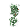
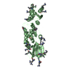
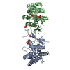
- Assembly
| Deposited unit | 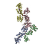
| ||||||||
|---|---|---|---|---|---|---|---|---|---|
| 1 | 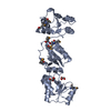
| ||||||||
| 2 | 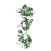
| ||||||||
| 3 | 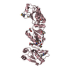
| ||||||||
| 4 | 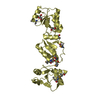
| ||||||||
| Unit cell |
|
-Components
| #1: Protein | Mass: 34313.457 Da / Num. of mol.: 4 / Fragment: BRCT 0,1 AND 2, RESIDUES 1-290 Source method: isolated from a genetically manipulated source Source: (gene. exp.) HOMO SAPIENS (human) / Plasmid: POPINH8 AND PGEX6P1 / Production host:  #2: Chemical | ChemComp-GOL /   Mass: 92.094 Da / Num. of mol.: 11 / Source method: obtained synthetically / Formula: C3H8O3 Mass: 92.094 Da / Num. of mol.: 11 / Source method: obtained synthetically / Formula: C3H8O3#3: Water | ChemComp-HOH / |  Mass: 18.015 Da / Num. of mol.: 340 / Source method: isolated from a natural source / Formula: H2O Mass: 18.015 Da / Num. of mol.: 340 / Source method: isolated from a natural source / Formula: H2OHas protein modification | Y | |
|---|
-Experimental details
-Experiment
| Experiment | Method: X-RAY DIFFRACTION / Number of used crystals: 1 |
|---|
- Sample preparation
| Crystal | Density Matthews: 2.58 Å3/Da / Density % sol: 0.52 % / Description: NONE |
|---|---|
| Crystal grow | pH: 7.5 Details: 0.1 M TRIS-HCL PH 7.5, 0.4 M MGCL2, 25% PEG 4000, 4% GLYCEROL, 0.01 M SPERMIDINE TETRA-HCL |
-Data collection
| Diffraction | Mean temperature: 100 K |
|---|---|
| Diffraction source | Source: SYNCHROTRON / Site: ESRF  / Beamline: ID14-4 / Wavelength: 0.9795 / Beamline: ID14-4 / Wavelength: 0.9795 |
| Detector | Type: ADSC CCD / Detector: CCD / Date: Jun 21, 2009 / Details: MIRRORS |
| Radiation | Protocol: SINGLE WAVELENGTH / Monochromatic (M) / Laue (L): M / Scattering type: x-ray |
| Radiation wavelength | Wavelength: 0.9795 Å / Relative weight: 1 |
| Reflection | Resolution: 2.47→55.91 Å / Num. obs: 40485 / % possible obs: 97.2 % / Observed criterion σ(I): 0 / Redundancy: 2.63 % / Biso Wilson estimate: 45.09 Å2 / Rmerge(I) obs: 0.1 / Net I/σ(I): 4.96 |
| Reflection shell | Resolution: 2.6→2.74 Å / Redundancy: 2.57 % / Rmerge(I) obs: 0.38 / Mean I/σ(I) obs: 1.99 / % possible all: 98.8 |
- Processing
| Software |
| |||||||||||||||||||||||||||||||||||||||||||||||||||||||||||||||||||||||||||||||||||||||||||||||||||||||||||||||||||||||||||||||||||||||||||||||||||||||||||||||||||||||||||||||||||||||||||||||||||||||||||||||||||||||||||||||||||||||||||||||||||||||||||||||||||||||||||||||||||||||||||||||||||||||||||||||||||||||||||||||||||||
|---|---|---|---|---|---|---|---|---|---|---|---|---|---|---|---|---|---|---|---|---|---|---|---|---|---|---|---|---|---|---|---|---|---|---|---|---|---|---|---|---|---|---|---|---|---|---|---|---|---|---|---|---|---|---|---|---|---|---|---|---|---|---|---|---|---|---|---|---|---|---|---|---|---|---|---|---|---|---|---|---|---|---|---|---|---|---|---|---|---|---|---|---|---|---|---|---|---|---|---|---|---|---|---|---|---|---|---|---|---|---|---|---|---|---|---|---|---|---|---|---|---|---|---|---|---|---|---|---|---|---|---|---|---|---|---|---|---|---|---|---|---|---|---|---|---|---|---|---|---|---|---|---|---|---|---|---|---|---|---|---|---|---|---|---|---|---|---|---|---|---|---|---|---|---|---|---|---|---|---|---|---|---|---|---|---|---|---|---|---|---|---|---|---|---|---|---|---|---|---|---|---|---|---|---|---|---|---|---|---|---|---|---|---|---|---|---|---|---|---|---|---|---|---|---|---|---|---|---|---|---|---|---|---|---|---|---|---|---|---|---|---|---|---|---|---|---|---|---|---|---|---|---|---|---|---|---|---|---|---|---|---|---|---|---|---|---|---|---|---|---|---|---|---|---|---|---|---|---|---|---|---|---|---|---|---|---|---|---|---|---|---|---|---|---|---|---|---|---|---|---|---|---|---|---|---|---|---|---|---|---|---|---|---|---|---|---|---|---|---|---|---|---|---|---|---|---|
| Refinement | Method to determine structure: MOLECULAR REPLACEMENT Starting model: PDB ENTRY 2XNH Resolution: 2.6→51.347 Å / SU ML: 0.92 / σ(F): 0.98 / Phase error: 29.4 / Stereochemistry target values: ML Details: DURING REFINEMENT UNMERGED REFLECTIONS (APPROXIMATELY DOUBLE THE NUMBER OF THE MERGED REFLECTIONS) IN ORDE TO TAKE INTO ACCOUNT THE ANOMALOUS CONTRIBUTIONS OF THE SEMET AT THE WAVELENGTH AT ...Details: DURING REFINEMENT UNMERGED REFLECTIONS (APPROXIMATELY DOUBLE THE NUMBER OF THE MERGED REFLECTIONS) IN ORDE TO TAKE INTO ACCOUNT THE ANOMALOUS CONTRIBUTIONS OF THE SEMET AT THE WAVELENGTH AT WHICH THE DATA SET WAS COLLECTED (WHICH HAPPENS TO BE THE INFLECTION POINT OF THE SE ANOMALOUS PEAK)
| |||||||||||||||||||||||||||||||||||||||||||||||||||||||||||||||||||||||||||||||||||||||||||||||||||||||||||||||||||||||||||||||||||||||||||||||||||||||||||||||||||||||||||||||||||||||||||||||||||||||||||||||||||||||||||||||||||||||||||||||||||||||||||||||||||||||||||||||||||||||||||||||||||||||||||||||||||||||||||||||||||||
| Solvent computation | Shrinkage radii: 0.9 Å / VDW probe radii: 1.11 Å / Solvent model: FLAT BULK SOLVENT MODEL / Bsol: 50.145 Å2 / ksol: 0.352 e/Å3 | |||||||||||||||||||||||||||||||||||||||||||||||||||||||||||||||||||||||||||||||||||||||||||||||||||||||||||||||||||||||||||||||||||||||||||||||||||||||||||||||||||||||||||||||||||||||||||||||||||||||||||||||||||||||||||||||||||||||||||||||||||||||||||||||||||||||||||||||||||||||||||||||||||||||||||||||||||||||||||||||||||||
| Displacement parameters | Biso mean: 57.5 Å2
| |||||||||||||||||||||||||||||||||||||||||||||||||||||||||||||||||||||||||||||||||||||||||||||||||||||||||||||||||||||||||||||||||||||||||||||||||||||||||||||||||||||||||||||||||||||||||||||||||||||||||||||||||||||||||||||||||||||||||||||||||||||||||||||||||||||||||||||||||||||||||||||||||||||||||||||||||||||||||||||||||||||
| Refinement step | Cycle: LAST / Resolution: 2.6→51.347 Å
| |||||||||||||||||||||||||||||||||||||||||||||||||||||||||||||||||||||||||||||||||||||||||||||||||||||||||||||||||||||||||||||||||||||||||||||||||||||||||||||||||||||||||||||||||||||||||||||||||||||||||||||||||||||||||||||||||||||||||||||||||||||||||||||||||||||||||||||||||||||||||||||||||||||||||||||||||||||||||||||||||||||
| Refine LS restraints |
| |||||||||||||||||||||||||||||||||||||||||||||||||||||||||||||||||||||||||||||||||||||||||||||||||||||||||||||||||||||||||||||||||||||||||||||||||||||||||||||||||||||||||||||||||||||||||||||||||||||||||||||||||||||||||||||||||||||||||||||||||||||||||||||||||||||||||||||||||||||||||||||||||||||||||||||||||||||||||||||||||||||
| LS refinement shell |
| |||||||||||||||||||||||||||||||||||||||||||||||||||||||||||||||||||||||||||||||||||||||||||||||||||||||||||||||||||||||||||||||||||||||||||||||||||||||||||||||||||||||||||||||||||||||||||||||||||||||||||||||||||||||||||||||||||||||||||||||||||||||||||||||||||||||||||||||||||||||||||||||||||||||||||||||||||||||||||||||||||||
| Refinement TLS params. | Method: refined / Refine-ID: X-RAY DIFFRACTION
| |||||||||||||||||||||||||||||||||||||||||||||||||||||||||||||||||||||||||||||||||||||||||||||||||||||||||||||||||||||||||||||||||||||||||||||||||||||||||||||||||||||||||||||||||||||||||||||||||||||||||||||||||||||||||||||||||||||||||||||||||||||||||||||||||||||||||||||||||||||||||||||||||||||||||||||||||||||||||||||||||||||
| Refinement TLS group |
|
