[English] 日本語
Yorodumi
- PDB-2xnh: Structure and function of the Rad9-binding region of the DNA dama... -

+
Open data


ID or keywords:

Loading...

-
Basic information

Entry
Database: PDB / ID: 2xnh
Title
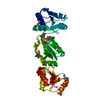
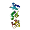
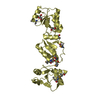
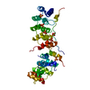
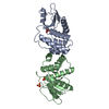
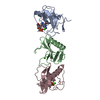
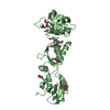
Structure and function of the Rad9-binding region of the DNA damage checkpoint adaptor TopBP1
ComponentsDNA TOPOISOMERASE 2-BINDING PROTEIN 1
KeywordsISOMERASE / PHOSPHORYLATION / PROTEIN-PROTEIN INTERACTION / DNA REPAIR
Function / homology
Function and homology information


BRCA1-B complex / phosphorylation-dependent protein binding / chromatin-protein adaptor activity / homologous recombination / protein localization to site of double-strand break / DNA replication checkpoint signaling / double-strand break repair via alternative nonhomologous end joining / double-strand break repair via classical nonhomologous end joining / mitotic DNA replication checkpoint signaling / HDR through Single Strand Annealing (SSA) ...BRCA1-B complex / phosphorylation-dependent protein binding / chromatin-protein adaptor activity / homologous recombination / protein localization to site of double-strand break / DNA replication checkpoint signaling / double-strand break repair via alternative nonhomologous end joining / double-strand break repair via classical nonhomologous end joining / mitotic DNA replication checkpoint signaling / HDR through Single Strand Annealing (SSA) / DNA metabolic process / Impaired BRCA2 binding to RAD51 / response to ionizing radiation / site of DNA damage / mitotic G2 DNA damage checkpoint signaling / Presynaptic phase of homologous DNA pairing and strand exchange / DNA replication initiation / chromosome organization / protein serine/threonine kinase activator activity / DNA damage checkpoint signaling / condensed nuclear chromosome / male germ cell nucleus / double-strand break repair via homologous recombination / G2/M DNA damage checkpoint / PML body / spindle pole / actin cytoskeleton / site of double-strand break / chromosome / Processing of DNA double-strand break ends / Regulation of TP53 Activity through Phosphorylation / nuclear body / DNA repair / centrosome / DNA damage response / DNA binding / nucleoplasm / identical protein binding / nucleus / plasma membrane / cytoplasm
Similarity search - Function
: / TopBP1, BRCT0 domain / TopBP1, first BRCT domain / Secretoglobin superfamily / twin BRCT domain / BRCT domain / BRCA1 C Terminus (BRCT) domain / breast cancer carboxy-terminal domain / BRCT domain profile. / BRCT domain ...: / TopBP1, BRCT0 domain / TopBP1, first BRCT domain / Secretoglobin superfamily / twin BRCT domain / BRCT domain / BRCA1 C Terminus (BRCT) domain / breast cancer carboxy-terminal domain / BRCT domain profile. / BRCT domain / BRCT domain superfamily / Rossmann fold / 3-Layer(aba) Sandwich / Alpha Beta
Similarity search - Domain/homology
IODIDE ION / DNA topoisomerase 2-binding protein 1
Similarity search - Component
Biological speciesHOMO SAPIENS (human)
MethodX-RAY DIFFRACTION / SYNCHROTRON / MAD / Resolution: 2.8 Å
AuthorsRappas, M. / Oliver, A.W. / Pearl, L.H.
CitationJournal: Nucleic Acids Res. / Year: 2011
Title: Structure and Function of the Rad9-Binding Region of the DNA-Damage Checkpoint Adaptor Topbp1.
Authors: Rappas, M. / Oliver, A.W. / Pearl, L.H.
History
DepositionAug 2, 2010Deposition site: PDBE / Processing site: PDBE
Revision 1.0Sep 1, 2010Provider: repository / Type: Initial release
Revision 1.1May 8, 2011Group: Version format compliance
Revision 1.2Jul 13, 2011Group: Version format compliance

-
Structure visualization

Structure viewerMolecule:
MolmilJmol/JSmol

Downloads & links

-
Assembly

Deposited unit
A: DNA TOPOISOMERASE 2-BINDING PROTEIN 1
hetero molecules


Theoretical massNumber of molelcules
Total (without water)36,01918
Polymers33,8621
Non-polymers2,15717
Water1,27971
1


  • Idetical with deposited unit
  • defined by author&software
TypeNameSymmetry operationNumber
identity operation1_555x,y,z1
MethodPISA
Unit cell
Length a, b, c (Å)115.770, 115.770, 115.770
Angle α, β, γ (deg.)90.00, 90.00, 90.00
Int Tables number198
Space group name H-MP213
Components on special symmetry positions
IDModelComponents
11A-1293-

IOD

21A-1302-

IOD

-
Components

#1: Protein DNA TOPOISOMERASE 2-BINDING PROTEIN 1 / DNA TOPOISOMERASE II-BINDING PROTEIN 1 / DNA TOPOISOMERASE II-BETA-BINDING PROTEIN 1 / TOPBP1


Mass: 33861.984 Da / Num. of mol.: 1 / Fragment: BRCT 0,1 AND 2, RESIDUES 1-287
Source method: isolated from a genetically manipulated source
Source: (gene. exp.) HOMO SAPIENS (human) / Plasmid: POPINH8, PGEX6P1 / Production host: ESCHERICHIA COLI (E. coli) / Strain (production host): ROSETTA2(DE3)PLYSS / References: UniProt: Q92547
#2: Chemical
ChemComp-IOD / IODIDE ION / Iodide


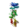
Mass: 126.904 Da / Num. of mol.: 17 / Source method: obtained synthetically / Formula: I
#3: Water ChemComp-HOH / water / Water


Mass: 18.015 Da / Num. of mol.: 71 / Source method: isolated from a natural source / Formula: H2O

-
Experimental details

-
Experiment

ExperimentMethod: X-RAY DIFFRACTION / Number of used crystals: 1

-
Sample preparation

CrystalDensity Matthews: 3.84 Å3/Da / Density % sol: 0.68 % / Description: NONE
Crystal growpH: 6.8
Details: 0.1M TRIS-HCL PH 6.8, 0.5M KI, 5% (V/V) GLYCEROL, 22.5% (W/V), 10% PEG 3350

-
Data collection

DiffractionMean temperature: 100 K
Diffraction sourceSource: SYNCHROTRON / Site: ESRF / Beamline: ID14-4 / Wavelength: 0.9795
DetectorType: ADSC CCD / Detector: CCD / Date: Jun 20, 2009 / Details: MIRRORS
RadiationProtocol: MAD / Monochromatic (M) / Laue (L): M / Scattering type: x-ray
Radiation wavelengthWavelength: 0.9795 Å / Relative weight: 1
ReflectionResolution: 2.6→51.77 Å / Num. obs: 13035 / % possible obs: 99.9 % / Observed criterion σ(I): 0 / Redundancy: 5.93 % / Biso Wilson estimate: 54.82 Å2 / Rmerge(I) obs: 0.09 / Net I/σ(I): 6.9
Reflection shellResolution: 2.8→2.95 Å / Redundancy: 6.09 % / Rmerge(I) obs: 0.5 / Mean I/σ(I) obs: 1.52 / % possible all: 100

-
Processing

Software
NameVersionClassification
PHENIX(PHENIX.REFINE)refinement
MOSFLMdata reduction
SCALAdata scaling
SHELXphasing
RefinementMethod to determine structure: MAD
Starting model: NONE

Resolution: 2.8→47.263 Å / SU ML: 1.16 / σ(F): 1.44 / Phase error: 23.97 / Stereochemistry target values: ML
RfactorNum. reflection% reflection
Rfree0.2513 1254 5.1 %
Rwork0.2318 --
obs0.2327 24687 99.83 %
Solvent computationShrinkage radii: 0.9 Å / VDW probe radii: 1.11 Å / Solvent model: FLAT BULK SOLVENT MODEL / Bsol: 58.687 Å2 / ksol: 0.339 e/Å3
Displacement parametersBiso mean: 58.7 Å2
Baniso -1Baniso -2Baniso -3
1-0 Å20 Å20 Å2
2--0 Å2-0 Å2
3---0 Å2
Refinement stepCycle: LAST / Resolution: 2.8→47.263 Å
ProteinNucleic acidLigandSolventTotal
Num. atoms2280 0 17 71 2368
Refine LS restraints
Refine-IDTypeDev idealNumber
X-RAY DIFFRACTIONf_bond_d0.0032330
X-RAY DIFFRACTIONf_angle_d0.6033135
X-RAY DIFFRACTIONf_dihedral_angle_d14.706875
X-RAY DIFFRACTIONf_chiral_restr0.041342
X-RAY DIFFRACTIONf_plane_restr0.002395
LS refinement shell
Resolution (Å)Rfactor RfreeNum. reflection RfreeRfactor RworkNum. reflection RworkRefine-ID% reflection obs (%)
2.8004-2.91250.29231470.28372648X-RAY DIFFRACTION100
2.9125-3.04510.3071500.27332592X-RAY DIFFRACTION100
3.0451-3.20560.30631280.26482606X-RAY DIFFRACTION100
3.2056-3.40640.28211240.24332609X-RAY DIFFRACTION100
3.4064-3.66930.24991410.21412584X-RAY DIFFRACTION100
3.6693-4.03840.23381500.19362596X-RAY DIFFRACTION100
4.0384-4.62230.21281440.17682605X-RAY DIFFRACTION100
4.6223-5.82190.17021340.19742603X-RAY DIFFRACTION100
5.8219-47.26960.25621360.24982590X-RAY DIFFRACTION99
Refinement TLS params.

Method: refined / Refine-ID: X-RAY DIFFRACTION

IDL112)L122)L132)L222)L232)L332)S11 (Å °)S12 (Å °)S13 (Å °)S21 (Å °)S22 (Å °)S23 (Å °)S31 (Å °)S32 (Å °)S33 (Å °)T112)T122)T132)T222)T232)T332)Origin x (Å)Origin y (Å)Origin z (Å)
11.2581-1.43521.33632.2667-2.49321.83480.1374-0.1004-0.4722-0.1710.30790.50650.1832-0.2714-0.38690.26010.021-0.1510.20270.0840.486536.792267.445576.9068
22.0077-2.15880.38812.7689-0.15280.1099-0.01980.2025-0.2981-0.1590.0630.12710.0064-0.1-0.00870.21740.0376-0.06740.2115-0.05680.128437.616889.142762.3073
30.7715-1.87931.29282.7296-0.66650.71230.18820.31530.0594-0.7324-0.26370.04430.0151-0.05270.04780.45330.1007-0.02330.32670.07930.18333.6415115.511850.837
Refinement TLS group
IDRefine-IDRefine TLS-IDSelection details
1X-RAY DIFFRACTION1CHAIN A AND RESID 6:103
2X-RAY DIFFRACTION2CHAIN A AND RESID 104:198
3X-RAY DIFFRACTION3CHAIN A AND RESID 199:287

+
About Yorodumi

-
News

-
Feb 9, 2022. New format data for meta-information of EMDB entries

New format data for meta-information of EMDB entries

  • Version 3 of the EMDB header file is now the official format.
  • The previous official version 1.9 will be removed from the archive.

Related info.:EMDB header

External links:wwPDB to switch to version 3 of the EMDB data model

-
Aug 12, 2020. Covid-19 info

Covid-19 info

URL: https://pdbj.org/emnavi/covid19.php

New page: Covid-19 featured information page in EM Navigator.

Related info.:Covid-19 info / Mar 5, 2020. Novel coronavirus structure data

+
Mar 5, 2020. Novel coronavirus structure data

Novel coronavirus structure data

Related info.:Yorodumi Speices / Aug 12, 2020. Covid-19 info

External links:COVID-19 featured content - PDBj / Molecule of the Month (242):Coronavirus Proteases

+
Jan 31, 2019. EMDB accession codes are about to change! (news from PDBe EMDB page)

EMDB accession codes are about to change! (news from PDBe EMDB page)

  • The allocation of 4 digits for EMDB accession codes will soon come to an end. Whilst these codes will remain in use, new EMDB accession codes will include an additional digit and will expand incrementally as the available range of codes is exhausted. The current 4-digit format prefixed with “EMD-” (i.e. EMD-XXXX) will advance to a 5-digit format (i.e. EMD-XXXXX), and so on. It is currently estimated that the 4-digit codes will be depleted around Spring 2019, at which point the 5-digit format will come into force.
  • The EM Navigator/Yorodumi systems omit the EMD- prefix.

Related info.:Q: What is EMD? / ID/Accession-code notation in Yorodumi/EM Navigator

External links:EMDB Accession Codes are Changing Soon! / Contact to PDBj

+
Jul 12, 2017. Major update of PDB

Major update of PDB

  • wwPDB released updated PDB data conforming to the new PDBx/mmCIF dictionary.
  • This is a major update changing the version number from 4 to 5, and with Remediation, in which all the entries are updated.
  • In this update, many items about electron microscopy experimental information are reorganized (e.g. em_software).
  • Now, EM Navigator and Yorodumi are based on the updated data.

External links:wwPDB Remediation / Enriched Model Files Conforming to OneDep Data Standards Now Available in the PDB FTP Archive

-
Yorodumi

Thousand views of thousand structures

  • Yorodumi is a browser for structure data from EMDB, PDB, SASBDB, etc.
  • This page is also the successor to EM Navigator detail page, and also detail information page/front-end page for Omokage search.
  • The word "yorodu" (or yorozu) is an old Japanese word meaning "ten thousand". "mi" (miru) is to see.

Related info.:EMDB / PDB / SASBDB / Comparison of 3 databanks / Yorodumi Search / Aug 31, 2016. New EM Navigator & Yorodumi / Yorodumi Papers / Jmol/JSmol / Function and homology information / Changes in new EM Navigator and Yorodumi

Read more