[English] 日本語
Yorodumi
- PDB-4rly: Crystal Structure of AnkB Ankyrin Repeats (R1-R9) in Complex with... -

+
Open data


ID or keywords:

Loading...

-
Basic information

Entry
Database: PDB / ID: 4rly
Title
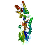
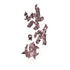
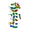
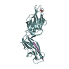
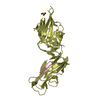
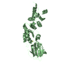
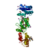
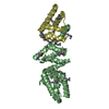
Crystal Structure of AnkB Ankyrin Repeats (R1-R9) in Complex with Nav1.2 Ankyrin Binding Domain
ComponentsNav1.2 - AnkB chimera
KeywordsSTRUCTURAL PROTEIN / ANK Repeat / Protein-protein interaction
Function / homology
Function and homology information


protein localization to T-tubule / atrial cardiac muscle cell to AV node cell communication / SA node cell to atrial cardiac muscle cell communication / regulation of calcium ion transmembrane transporter activity / positive regulation of calcium ion transmembrane transporter activity / protein localization to M-band / positive regulation of potassium ion transmembrane transporter activity / T-tubule organization / SA node cell action potential / membrane depolarization during SA node cell action potential ...protein localization to T-tubule / atrial cardiac muscle cell to AV node cell communication / SA node cell to atrial cardiac muscle cell communication / regulation of calcium ion transmembrane transporter activity / positive regulation of calcium ion transmembrane transporter activity / protein localization to M-band / positive regulation of potassium ion transmembrane transporter activity / T-tubule organization / SA node cell action potential / membrane depolarization during SA node cell action potential / protein localization to organelle / paranodal junction assembly / phosphorylation-dependent protein binding / regulation of SA node cell action potential / regulation of atrial cardiac muscle cell action potential / positive regulation of cation channel activity / atrial cardiac muscle cell action potential / protein localization to endoplasmic reticulum / sarcoplasmic reticulum calcium ion transport / intrinsic apoptotic signaling pathway in response to osmotic stress / nerve development / paranode region of axon / cytoskeletal anchor activity / atrial septum development / positive regulation of potassium ion transport / dentate gyrus development / node of Ranvier / ventricular cardiac muscle cell action potential / costamere / response to methylmercury / regulation of release of sequestered calcium ion into cytosol / positive regulation of calcium ion transport / cardiac muscle cell action potential involved in contraction / regulation of ventricular cardiac muscle cell membrane repolarization / M band / regulation of cardiac muscle cell contraction / regulation of cardiac muscle contraction by calcium ion signaling / protein localization to cell surface / voltage-gated sodium channel complex / membrane depolarization during action potential / Interaction between L1 and Ankyrins / A band / voltage-gated sodium channel activity / sodium ion transport / spectrin binding / Phase 0 - rapid depolarisation / regulation of heart rate by cardiac conduction / regulation of calcium ion transport / intercalated disc / regulation of cardiac muscle contraction / sodium ion transmembrane transport / neuronal action potential / regulation of cardiac muscle contraction by regulation of the release of sequestered calcium ion / COPI-mediated anterograde transport / T-tubule / myelination / regulation of heart rate / protein localization to plasma membrane / determination of adult lifespan / regulation of protein stability / protein localization / recycling endosome / structural constituent of cytoskeleton / sarcolemma / memory / Sensory perception of sweet, bitter, and umami (glutamate) taste / Z disc / intracellular calcium ion homeostasis / endocytosis / protein transport / protein-macromolecule adaptor activity / presynaptic membrane / nervous system development / ATPase binding / cellular response to hypoxia / basolateral plasma membrane / postsynaptic membrane / neuron apoptotic process / transmembrane transporter binding / lysosome / early endosome / cytoskeleton / protein stabilization / calmodulin binding / neuron projection / apical plasma membrane / axon / glutamatergic synapse / positive regulation of gene expression / protein kinase binding / enzyme binding / mitochondrion / membrane / plasma membrane / cytosol
Similarity search - Function
Ankyrin, UPA domain / UPA domain / Domain present in ZO-1 and Unc5-like netrin receptors / ZU5 domain / ZU5 domain / ZU5 domain profile. / Voltage-gated Na+ ion channel, cytoplasmic domain / Cytoplasmic domain of voltage-gated Na+ ion channel / Voltage-gated sodium channel alpha subunit, inactivation gate / Sodium ion transport-associated ...Ankyrin, UPA domain / UPA domain / Domain present in ZO-1 and Unc5-like netrin receptors / ZU5 domain / ZU5 domain / ZU5 domain profile. / Voltage-gated Na+ ion channel, cytoplasmic domain / Cytoplasmic domain of voltage-gated Na+ ion channel / Voltage-gated sodium channel alpha subunit, inactivation gate / Sodium ion transport-associated / Sodium ion transport-associated / Voltage gated sodium channel, alpha subunit / Death domain profile. / Voltage-gated cation channel calcium and sodium / DEATH domain, found in proteins involved in cell death (apoptosis). / Death domain / Death domain / Ankyrin repeat-containing domain / Ankyrin repeats (many copies) / Short calmodulin-binding motif containing conserved Ile and Gln residues. / IQ motif profile. / IQ motif, EF-hand binding site / Voltage-dependent channel domain superfamily / Death-like domain superfamily / Ankyrin repeat / Ankyrin repeats (3 copies) / Ankyrin repeat profile. / Ankyrin repeat region circular profile. / ankyrin repeats / Ankyrin repeat / Ankyrin repeat-containing domain superfamily / Serine Threonine Protein Phosphatase 5, Tetratricopeptide repeat / Ion transport domain / Ion transport protein / Alpha Horseshoe / Mainly Alpha
Similarity search - Domain/homology
Sodium channel Nav1.2 / Ankyrin-2 / Sodium channel protein type 2 subunit alpha
Similarity search - Component
Biological speciesMus musculus (house mouse)
Homo sapiens (human)
MethodX-RAY DIFFRACTION / SYNCHROTRON / MOLECULAR REPLACEMENT / molecular replacement / Resolution: 2.5032 Å
AuthorsWei, Z. / Wang, C. / Zhang, M.
CitationJournal: Elife / Year: 2014
Title: Structural basis of diverse membrane target recognitions by ankyrins.
Authors: Wang, C. / Wei, Z. / Chen, K. / Ye, F. / Yu, C. / Bennett, V. / Zhang, M.
History
DepositionOct 18, 2014Deposition site: RCSB / Processing site: RCSB
Revision 1.0Nov 26, 2014Provider: repository / Type: Initial release
Revision 1.1Jan 14, 2015Group: Refinement description
Revision 1.2May 20, 2015Group: Database references
Revision 1.3Aug 2, 2017Group: Refinement description / Source and taxonomy / Category: entity_src_gen / software
Revision 1.4Nov 22, 2017Group: Refinement description / Category: software
Revision 1.5Feb 28, 2024Group: Data collection / Database references / Derived calculations
Category: chem_comp_atom / chem_comp_bond ...chem_comp_atom / chem_comp_bond / database_2 / struct_ref_seq_dif / struct_site
Item: _database_2.pdbx_DOI / _database_2.pdbx_database_accession ..._database_2.pdbx_DOI / _database_2.pdbx_database_accession / _struct_ref_seq_dif.details / _struct_site.pdbx_auth_asym_id / _struct_site.pdbx_auth_comp_id / _struct_site.pdbx_auth_seq_id

-
Structure visualization

Structure viewerMolecule:
MolmilJmol/JSmol

Downloads & links

-
Assembly

Deposited unit
A: Nav1.2 - AnkB chimera
hetero molecules


Theoretical massNumber of molelcules
Total (without water)35,4838
Polymers34,8101
Non-polymers6727
Water1,33374
1


  • Idetical with deposited unit
  • defined by author
TypeNameSymmetry operationNumber
identity operation1_555x,y,z1
2
A: Nav1.2 - AnkB chimera
hetero molecules

A: Nav1.2 - AnkB chimera
hetero molecules


Theoretical massNumber of molelcules
Total (without water)70,96516
Polymers69,6202
Non-polymers1,34514
Water362
TypeNameSymmetry operationNumber
identity operation1_555x,y,z1
crystal symmetry operation6_556x,-y,-z+11
Buried area2460 Å2
ΔGint-7 kcal/mol
Surface area26500 Å2
MethodPISA
Unit cell
Length a, b, c (Å)102.295, 102.295, 106.008
Angle α, β, γ (deg.)90.000, 90.000, 90.000
Int Tables number93
Space group name H-MP4222
Components on special symmetry positions
IDModelComponents
11A-2403-

SO4

-
Components

#1: Protein Nav1.2 - AnkB chimera


Mass: 34810.133 Da / Num. of mol.: 1
Source method: isolated from a genetically manipulated source
Source: (gene. exp.) Mus musculus (house mouse), (gene. exp.) Homo sapiens (human)
Gene: ANK2, Scn2a, Scn2a1
Plasmid details: The Nav1.2 Ankyrin binding domain is fused to the N-terminus of AnkB Ankyrin re peats
Plasmid: pET32m / Production host: Escherichia coli (E. coli) / Strain (production host): BL21(DE3)
References: UniProt: A9JQD3, UniProt: Q01484, UniProt: Q99250*PLUS
#2: Chemical
ChemComp-SO4 / SULFATE ION


Mass: 96.063 Da / Num. of mol.: 7 / Source method: obtained synthetically / Formula: SO4
#3: Water ChemComp-HOH / water


Mass: 18.015 Da / Num. of mol.: 74 / Source method: isolated from a natural source / Formula: H2O

-
Experimental details

-
Experiment

ExperimentMethod: X-RAY DIFFRACTION / Number of used crystals: 1

-
Sample preparation

CrystalDensity Matthews: 3.98 Å3/Da / Density % sol: 69.12 %
Crystal growTemperature: 289 K / Method: vapor diffusion, hanging drop / pH: 6.5
Details: 1.8 M ammonium sulfate, 6-8% dioxane, and 0.1 M MES , pH 6.5, VAPOR DIFFUSION, HANGING DROP, temperature 289K

-
Data collection

DiffractionMean temperature: 100 K
Diffraction sourceSource: SYNCHROTRON / Site: SSRF / Beamline: BL17U
DetectorType: ADSC QUANTUM 315r / Detector: CCD / Date: Jun 1, 2012
RadiationProtocol: SINGLE WAVELENGTH / Monochromatic (M) / Laue (L): M / Scattering type: x-ray
Radiation wavelengthRelative weight: 1
ReflectionResolution: 2.5→50 Å / Num. obs: 19914 / % possible obs: 99.4 % / Redundancy: 9.5 % / Biso Wilson estimate: 53.21 Å2 / Rmerge(I) obs: 0.077 / Χ2: 1.209 / Net I/σ(I): 11.8
Reflection shell
Resolution (Å)Redundancy (%)Rmerge(I) obsNum. unique allΧ2Diffraction-ID% possible all
2.5-2.549.70.7489771.0551100
2.54-2.599.80.6159821.056199.8
2.59-2.649.70.5369601.086199.8
2.64-2.699.60.4529771.098199.9
2.69-2.759.80.3819781.1441100
2.75-2.829.70.3439851.104199.9
2.82-2.899.70.2869971.199199.8
2.89-2.969.60.249721.209199.9
2.96-3.059.70.199781.279199.9
3.05-3.159.70.15110041.308199.9
3.15-3.269.70.1219891.2781100
3.26-3.399.60.0969851.2971100
3.39-3.559.50.07810101.2241100
3.55-3.739.50.0649831.31100
3.73-3.979.40.05510051.161199.8
3.97-4.279.20.05710181.329199.7
4.27-4.79.10.05310011.309199.3
4.7-5.388.90.05610241.277199
5.38-6.788.80.04710311.175198.5
6.78-508.80.03910581.297193.2

-
Phasing

PhasingMethod: molecular replacement

-
Processing

Software
NameVersionClassificationNB
DENZOdata reduction
SCALEPACKdata scaling
PHASERphasing
PHENIXrefinement
PDB_EXTRACT3.15data extraction
ADSCQuantumdata collection
HKL-2000data reduction
HKL-2000data scaling
RefinementMethod to determine structure: MOLECULAR REPLACEMENT / Resolution: 2.5032→34.229 Å / FOM work R set: 0.823 / SU ML: 0.65 / σ(F): 1.34 / Phase error: 23.7 / Stereochemistry target values: ML
RfactorNum. reflection% reflection
Rfree0.2384 1009 5.09 %
Rwork0.1875 --
obs0.19 19832 99.16 %
Solvent computationShrinkage radii: 0.83 Å / VDW probe radii: 1.1 Å / Solvent model: FLAT BULK SOLVENT MODEL / Bsol: 57.211 Å2 / ksol: 0.341 e/Å3
Displacement parametersBiso max: 163.12 Å2 / Biso mean: 69.27 Å2 / Biso min: 31.77 Å2
Baniso -1Baniso -2Baniso -3
1-10.595 Å20 Å2-0 Å2
2--10.595 Å2-0 Å2
3----21.19 Å2
Refinement stepCycle: LAST / Resolution: 2.5032→34.229 Å
ProteinNucleic acidLigandSolventTotal
Num. atoms2262 0 35 74 2371
Refine LS restraints
Refine-IDTypeDev idealNumber
X-RAY DIFFRACTIONf_bond_d0.0152318
X-RAY DIFFRACTIONf_angle_d1.5433151
X-RAY DIFFRACTIONf_chiral_restr0.098380
X-RAY DIFFRACTIONf_plane_restr0.006410
X-RAY DIFFRACTIONf_dihedral_angle_d17.041811
LS refinement shell

Refine-ID: X-RAY DIFFRACTION / Total num. of bins used: 7

Resolution (Å)Rfactor RfreeNum. reflection RfreeRfactor RworkNum. reflection RworkNum. reflection all% reflection obs (%)
2.5032-2.63510.24531490.227226292778100
2.6351-2.80010.29741570.228726372794100
2.8001-3.01620.29781420.230226652807100
3.0162-3.31950.26711560.199726592815100
3.3195-3.79930.24241340.184927122846100
3.7993-4.78460.20211370.15322717285499
4.7846-34.23260.22261340.18772804293896
Refinement TLS params.

Method: refined / Refine-ID: X-RAY DIFFRACTION

IDL112)L122)L132)L222)L232)L332)S11 (Å °)S12 (Å °)S13 (Å °)S21 (Å °)S22 (Å °)S23 (Å °)S31 (Å °)S32 (Å °)S33 (Å °)T112)T122)T132)T222)T232)T332)Origin x (Å)Origin y (Å)Origin z (Å)
12.2584-0.98240.76961.5564-0.28110.65230.0283-0.6322-0.3590.69070.02620.29610.1587-0.15440.04990.4383-0.03050.00640.30590.040.445340.9904-23.526358.6885
20.3395-0.37270.44680.5096-0.32230.46190.00760.29310.0042-0.27230.01250.00480.3585-0.146-0.00020.34270.0031-0.01160.4506-0.06230.457737.4855-12.940943.2483
30.2332-0.18790.65441.59511.18053.66710.4860.17170.30650.0757-0.62650.50430.288-1.1529-0.04770.30850.05440.02220.52350.0260.477834.4303-0.606736.9452
40.44260.21990.09640.53280.02720.02040.33270.5707-0.0248-0.1411-0.13510.24241.33440.426-0.01460.84820.2515-0.01210.63810.03790.433134.66171.896225.3009
50.8728-0.00940.79570.3579-1.03524.4035-0.0682-0.24230.30220.6543-0.1773-0.0859-1.0005-0.1046-0.15220.4230.093-0.03350.61240.03550.409929.334415.132530.8628
60.4877-0.30610.23760.6346-0.32840.30820.23220.1950.1092-0.4589-0.1260.17710.13120.467-0.0010.63870.18490.06120.49060.02350.295226.178610.392319.7096
70.0238-0.0655-0.03880.0252-0.0040.08540.21350.5616-0.4993-0.8505-0.26520.41611.261-0.25740.00130.79710.1551-0.07890.6192-0.02410.362617.6745.72415.4184
80.4193-0.18090.370.62670.07350.58860.30970.1730.6768-1.13510.0144-0.3025-0.6460.84510.26491.04780.23880.01450.81820.02070.487619.46514.03757.3215
90.31010.1776-0.23611.1644-0.33780.23280.82450.4076-0.0819-0.372-0.2140.55970.692-0.05981.03071.51770.1229-0.20360.913-0.2350.357713.23233.64356.8579
102.0331-0.0088-0.14311.8716-1.20582.7395-0.326-0.23160.04620.8038-0.0654-0.3871-0.4753-0.3354-0.11170.57240.02410.05720.4609-0.02780.424434.9676-4.633657.1305
Refinement TLS group
IDRefine-IDRefine TLS-IDSelection detailsAuth asym-IDAuth seq-ID
1X-RAY DIFFRACTION1chain 'A' and (resseq 2029:2086)A2029 - 2086
2X-RAY DIFFRACTION2chain 'A' and (resseq 2087:2152)A2087 - 2152
3X-RAY DIFFRACTION3chain 'A' and (resseq 2153:2183)A2153 - 2183
4X-RAY DIFFRACTION4chain 'A' and (resseq 2184:2204)A2184 - 2204
5X-RAY DIFFRACTION5chain 'A' and (resseq 2205:2227)A2205 - 2227
6X-RAY DIFFRACTION6chain 'A' and (resseq 2228:2268)A2228 - 2268
7X-RAY DIFFRACTION7chain 'A' and (resseq 2269:2287)A2269 - 2287
8X-RAY DIFFRACTION8chain 'A' and (resseq 2288:2301)A2288 - 2301
9X-RAY DIFFRACTION9chain 'A' and (resseq 2302:2322)A2302 - 2322
10X-RAY DIFFRACTION10chain 'A' and (resseq 1113:1132)A1113 - 1132

+
About Yorodumi

-
News

-
Feb 9, 2022. New format data for meta-information of EMDB entries

New format data for meta-information of EMDB entries

  • Version 3 of the EMDB header file is now the official format.
  • The previous official version 1.9 will be removed from the archive.

Related info.:EMDB header

External links:wwPDB to switch to version 3 of the EMDB data model

-
Aug 12, 2020. Covid-19 info

Covid-19 info

URL: https://pdbjlvh1.pdbj.org/emnavi/covid19.php

New page: Covid-19 featured information page in EM Navigator.

Related info.:Covid-19 info / Mar 5, 2020. Novel coronavirus structure data

+
Mar 5, 2020. Novel coronavirus structure data

Novel coronavirus structure data

Related info.:Yorodumi Speices / Aug 12, 2020. Covid-19 info

External links:COVID-19 featured content - PDBj / Molecule of the Month (242):Coronavirus Proteases

+
Jan 31, 2019. EMDB accession codes are about to change! (news from PDBe EMDB page)

EMDB accession codes are about to change! (news from PDBe EMDB page)

  • The allocation of 4 digits for EMDB accession codes will soon come to an end. Whilst these codes will remain in use, new EMDB accession codes will include an additional digit and will expand incrementally as the available range of codes is exhausted. The current 4-digit format prefixed with “EMD-” (i.e. EMD-XXXX) will advance to a 5-digit format (i.e. EMD-XXXXX), and so on. It is currently estimated that the 4-digit codes will be depleted around Spring 2019, at which point the 5-digit format will come into force.
  • The EM Navigator/Yorodumi systems omit the EMD- prefix.

Related info.:Q: What is EMD? / ID/Accession-code notation in Yorodumi/EM Navigator

External links:EMDB Accession Codes are Changing Soon! / Contact to PDBj

+
Jul 12, 2017. Major update of PDB

Major update of PDB

  • wwPDB released updated PDB data conforming to the new PDBx/mmCIF dictionary.
  • This is a major update changing the version number from 4 to 5, and with Remediation, in which all the entries are updated.
  • In this update, many items about electron microscopy experimental information are reorganized (e.g. em_software).
  • Now, EM Navigator and Yorodumi are based on the updated data.

External links:wwPDB Remediation / Enriched Model Files Conforming to OneDep Data Standards Now Available in the PDB FTP Archive

-
Yorodumi

Thousand views of thousand structures

  • Yorodumi is a browser for structure data from EMDB, PDB, SASBDB, etc.
  • This page is also the successor to EM Navigator detail page, and also detail information page/front-end page for Omokage search.
  • The word "yorodu" (or yorozu) is an old Japanese word meaning "ten thousand". "mi" (miru) is to see.

Related info.:EMDB / PDB / SASBDB / Comparison of 3 databanks / Yorodumi Search / Aug 31, 2016. New EM Navigator & Yorodumi / Yorodumi Papers / Jmol/JSmol / Function and homology information / Changes in new EM Navigator and Yorodumi

Read more