- PDB-4n40: Crystal structure of human Epithelial cell-transforming sequence ... -
+
Open data
ID or keywords:
Loading...
-
Basic information
Entry
Database: PDB / ID: 4n40
Title
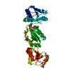
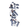
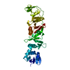
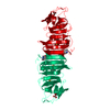
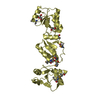
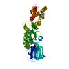
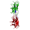
Crystal structure of human Epithelial cell-transforming sequence 2 protein
Components
Protein ECT2
Keywords
CELL CYCLE / triple brct domains
Function / homology
Function and homology information
regulation of cytokinesis, actomyosin contractile ring assembly / centralspindlin complex / positive regulation of mitotic cytokinetic process / regulation of protein kinase activity / activation of protein kinase activity / regulation of attachment of spindle microtubules to kinetochore / bicellular tight junction assembly / activation of GTPase activity / regulation of small GTPase mediated signal transduction / RHOB GTPase cycle ...regulation of cytokinesis, actomyosin contractile ring assembly / centralspindlin complex / positive regulation of mitotic cytokinetic process / regulation of protein kinase activity / activation of protein kinase activity / regulation of attachment of spindle microtubules to kinetochore / bicellular tight junction assembly / activation of GTPase activity / regulation of small GTPase mediated signal transduction / RHOB GTPase cycle / NRAGE signals death through JNK / positive regulation of cytokinesis / cleavage furrow / mitotic cytokinesis / CDC42 GTPase cycle / positive regulation of GTPase activity / RHOA GTPase cycle / bicellular tight junction / RAC1 GTPase cycle / positive regulation of neuron differentiation / guanyl-nucleotide exchange factor activity / cellular response to calcium ion / GTPase activator activity / cellular response to ionizing radiation / positive regulation of protein import into nucleus / protein homooligomerization / small GTPase binding / cellular response to hydrogen peroxide / cell-cell junction / mitotic spindle / nervous system development / protein transport / G alpha (12/13) signalling events / midbody / cell cortex / cell differentiation / positive regulation of canonical NF-kappaB signal transduction / intracellular signal transduction / positive regulation of apoptotic process / centrosome / protein homodimerization activity / nucleoplasm / nucleus / cytoplasm / cytosol Similarity search - Function
In the structure databanks used in Yorodumi, some data are registered as the other names, "COVID-19 virus" and "2019-nCoV". Here are the details of the virus and the list of structure data.
Jan 31, 2019. EMDB accession codes are about to change! (news from PDBe EMDB page)
EMDB accession codes are about to change! (news from PDBe EMDB page)
The allocation of 4 digits for EMDB accession codes will soon come to an end. Whilst these codes will remain in use, new EMDB accession codes will include an additional digit and will expand incrementally as the available range of codes is exhausted. The current 4-digit format prefixed with “EMD-” (i.e. EMD-XXXX) will advance to a 5-digit format (i.e. EMD-XXXXX), and so on. It is currently estimated that the 4-digit codes will be depleted around Spring 2019, at which point the 5-digit format will come into force.
The EM Navigator/Yorodumi systems omit the EMD- prefix.
Related info.:Q: What is EMD? / ID/Accession-code notation in Yorodumi/EM Navigator
Yorodumi is a browser for structure data from EMDB, PDB, SASBDB, etc.
This page is also the successor to EM Navigator detail page, and also detail information page/front-end page for Omokage search.
The word "yorodu" (or yorozu) is an old Japanese word meaning "ten thousand". "mi" (miru) is to see.
Related info.:EMDB / PDB / SASBDB / Comparison of 3 databanks / Yorodumi Search / Aug 31, 2016. New EM Navigator & Yorodumi / Yorodumi Papers / Jmol/JSmol / Function and homology information / Changes in new EM Navigator and Yorodumi
 Movie
Movie Controller

 Yorodumi
 Open data
 Basic information
 Components
 Keywords
 Function and homology information
 Homo sapiens (human)
 X-RAY DIFFRACTION /
  Authors
 Citation
 Structure visualization
 Downloads & links
 Other downloads
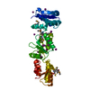
 PDBj
PDBj


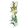
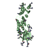
 Assembly
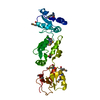


 Sample preparation
 Processing