 Movie
Movie Controller


+ Open data

- Basic information
| Entry | Database: PDB / ID: 7rwt | ||||||
|---|---|---|---|---|---|---|---|
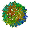
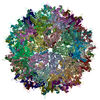
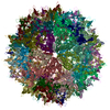
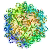
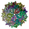
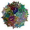
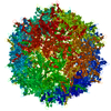
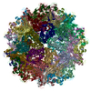
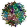
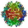
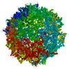
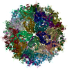
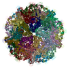
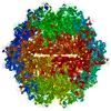
| Title | Adeno-associated virus type 2 | ||||||
 Components Components | Capsid protein VP1 | ||||||
 Keywords Keywords | VIRUS / Adeno-associated virus type 2 | ||||||
| Function / homology |  Function and homology information Function and homology informationsymbiont entry into host cell via permeabilization of host membrane / host cell nucleolus / T=1 icosahedral viral capsid / clathrin-dependent endocytosis of virus by host cell / virion attachment to host cell / structural molecule activity Similarity search - Function | ||||||
| Biological species |  Adeno-associated dependoparvovirus A Adeno-associated dependoparvovirus A | ||||||
| Method | ELECTRON MICROSCOPY / single particle reconstruction / cryo EM / Resolution: 2.43 Å | ||||||
 Authors Authors | Hull, J.A. / Mietzsch, M. / Chipman, P. / Strugatsky, D. / McKenna, R. | ||||||
| Funding support |  United States, 1items United States, 1items
| ||||||
 Citation Citation | Journal: Virology / Year: 2022 Title: Structural characterization of an envelope-associated adeno-associated virus type 2 capsid. Authors: Joshua A Hull / Mario Mietzsch / Paul Chipman / David Strugatsky / Robert McKenna / Abstract: Adeno-associated virus (AAV) are classified as non-enveloped ssDNA viruses. However, AAV capsids embedded within exosomes have been observed, and it has been suggested that the AAV membrane ...Adeno-associated virus (AAV) are classified as non-enveloped ssDNA viruses. However, AAV capsids embedded within exosomes have been observed, and it has been suggested that the AAV membrane associated accessory protein (MAAP) may play a role in envelope-associated AAV (EA-AAV) capsid formation. Here, we observed and selected sufficient homogeneous EA-AAV capsids of AAV2, produced using the Sf9 baculoviral expression system, to determine the cryo-electron microscopy (cryo-EM) structure at 3.14 Å resolution. The reconstructed map confirmed that the EA-AAV capsid, showed no significant structural variation compared to the non-envelope capsid. In addition, the Sf9 expression system used implies the notion that MAAP may enhance exosome AAV encapsulation. Furthermore, we speculate that these EA-AAV capsids may have therapeutic benefits over the currently used non-envelope AAV capsids, with advantages in immune evasion and/or improved infectivity. | ||||||
| History |
|
- Structure visualization
| Movie |
Movie viewer |
|---|---|
| Structure viewer | Molecule: MolmilJmol/JSmol |
- Downloads & links
-Download
| PDBx/mmCIF format | 7rwt.cif.gz | 5.7 MB | Display | PDBx/mmCIF format |
|---|---|---|---|---|
| PDB format | pdb7rwt.ent.gz | Display | PDB format | |
| PDBx/mmJSON format | 7rwt.json.gz | Tree view | PDBx/mmJSON format | |
| Others |  Other downloads Other downloads |
-Validation report
| Arichive directory | https://data.pdbj.org/pub/pdb/validation_reports/rw/7rwtftp://data.pdbj.org/pub/pdb/validation_reports/rw/7rwt | HTTPS FTP |
|---|
-Related structure data
| Related structure data | 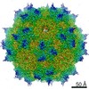 24719MC  7rwlC M: map data used to model this data C: citing same article ( |
|---|---|
| Similar structure data |


-Links
 PDBj
PDBj


- Assembly
| Deposited unit | 
|
|---|---|
| 1 |
|
-Components
| #1: Protein | Mass: 82031.352 Da / Num. of mol.: 60 Source method: isolated from a genetically manipulated source Source: (gene. exp.) Adeno-associated dependoparvovirus A / Gene: VP1 / Production host:   Spodoptera frugiperda (fall armyworm) / References: UniProt: P03135 Spodoptera frugiperda (fall armyworm) / References: UniProt: P03135 |
|---|
-Experimental details
-Experiment
| Experiment | Method: ELECTRON MICROSCOPY |
|---|---|
| EM experiment | Aggregation state: PARTICLE / 3D reconstruction method: single particle reconstruction |
- Sample preparation
| Component | Name: Adeno-associated dependoparvovirus A / Type: VIRUS / Entity ID: all / Source: RECOMBINANT |
|---|---|
| Source (natural) | Organism: Adeno-associated dependoparvovirus A |
| Source (recombinant) | Organism: Spodoptera frugiperda (fall armyworm) |
| Details of virus | Empty: YES / Enveloped: NO / Isolate: SEROTYPE / Type: VIRUS-LIKE PARTICLE |
| Virus shell | Triangulation number (T number): 1 |
| Buffer solution | pH: 7.4 |
| Specimen | Embedding applied: NO / Shadowing applied: NO / Staining applied: NO / Vitrification applied: YES |
| Vitrification | Cryogen name: ETHANE |
- Electron microscopy imaging
| Experimental equipment |  Model: Titan Krios / Image courtesy: FEI Company |
|---|---|
| Microscopy | Model: FEI TITAN KRIOS |
| Electron gun | Electron source:  FIELD EMISSION GUN / Accelerating voltage: 300 kV / Illumination mode: FLOOD BEAM FIELD EMISSION GUN / Accelerating voltage: 300 kV / Illumination mode: FLOOD BEAM |
| Electron lens | Mode: BRIGHT FIELD |
| Image recording | Electron dose: 34 e/Å2 / Film or detector model: GATAN K3 (6k x 4k) |
- Processing
| CTF correction | Type: PHASE FLIPPING AND AMPLITUDE CORRECTION |
|---|---|
| 3D reconstruction | Resolution: 2.43 Å / Resolution method: FSC 0.143 CUT-OFF / Num. of particles: 46289 / Symmetry type: POINT |
