 Movie
Movie Controller

[English] 日本語
 Yorodumi
- PDB-7rnv: SH2 domain of guanine nucleotide exchange factor Vav2 in complex ... -
+ Open data

- Basic information
| Entry | Database: PDB / ID: 7rnv | ||||||
|---|---|---|---|---|---|---|---|
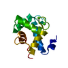
| Title | SH2 domain of guanine nucleotide exchange factor Vav2 in complex with an actin peptide with phosphorylated tyrosine 53 | ||||||
 Components Components |
| ||||||
 Keywords Keywords | PEPTIDE BINDING PROTEIN / phosphorylated tyrosine binding protein / actin peptide | ||||||
| Function / homology |  Function and homology information Function and homology informationimmune response-regulating cell surface receptor signaling pathway / Formation of the dystrophin-glycoprotein complex (DGC) / Striated Muscle Contraction / regulation of small GTPase mediated signal transduction / Azathioprine ADME / epidermal growth factor receptor binding / regulation of GTPase activity / lamellipodium assembly / RHOB GTPase cycle / NRAGE signals death through JNK ...immune response-regulating cell surface receptor signaling pathway / Formation of the dystrophin-glycoprotein complex (DGC) / Striated Muscle Contraction / regulation of small GTPase mediated signal transduction / Azathioprine ADME / epidermal growth factor receptor binding / regulation of GTPase activity / lamellipodium assembly / RHOB GTPase cycle / NRAGE signals death through JNK / small GTPase-mediated signal transduction / myosin binding / RHOC GTPase cycle / regulation of cell size / Fc-epsilon receptor signaling pathway / Fc-gamma receptor signaling pathway involved in phagocytosis / mesenchyme migration / CDC42 GTPase cycle / striated muscle thin filament / skeletal muscle thin filament assembly / RHOG GTPase cycle / EPH-ephrin mediated repulsion of cells / RHOA GTPase cycle / RAC2 GTPase cycle / RAC3 GTPase cycle / vascular endothelial growth factor receptor signaling pathway / skeletal muscle fiber development / GPVI-mediated activation cascade / stress fiber / phosphotyrosine residue binding / RAC1 GTPase cycle / FCERI mediated Ca+2 mobilization / guanyl-nucleotide exchange factor activity / muscle contraction / sarcomere / Signal transduction by L1 / VEGFR2 mediated vascular permeability / FCGR3A-mediated phagocytosis / actin filament / FCERI mediated MAPK activation / filopodium / ADP binding / platelet activation / Regulation of actin dynamics for phagocytic cup formation / structural constituent of cytoskeleton / VEGFA-VEGFR2 Pathway / Hydrolases; Acting on acid anhydrides; Acting on acid anhydrides to facilitate cellular and subcellular movement / cellular response to xenobiotic stimulus / DAP12 signaling / cell migration / actin cytoskeleton / G alpha (12/13) signalling events / lamellipodium / cell body / angiogenesis / blood microparticle / positive regulation of phosphatidylinositol 3-kinase/protein kinase B signal transduction / hydrolase activity / positive regulation of gene expression / signal transduction / extracellular space / extracellular exosome / zinc ion binding / ATP binding / plasma membrane / cytoplasm / cytosol Similarity search - Function | ||||||
| Biological species |  Homo sapiens (human) Homo sapiens (human) | ||||||
| Method |  X-RAY DIFFRACTION / MOLECULAR REPLACEMENT / Resolution: 2.15 Å X-RAY DIFFRACTION / MOLECULAR REPLACEMENT / Resolution: 2.15 Å | ||||||
 Authors Authors | Dai, S. / Horton, J.R. / Cheng, X. | ||||||
| Funding support |  United States, 1items United States, 1items
| ||||||
 Citation Citation | Journal: To Be Published Title: The Functional Analysis of a Major Tyrosine Phosphorylation Site on Actin Authors: Amelie, A. / Dai, S. / Shen, X. / Horton, J.R. / Zhang, X. / Cheng, X. | ||||||
| History |
|
- Structure visualization
| Structure viewer | Molecule: MolmilJmol/JSmol |
|---|
- Downloads & links
-Download
| PDBx/mmCIF format | 7rnv.cif.gz | 45.5 KB | Display | PDBx/mmCIF format |
|---|---|---|---|---|
| PDB format | pdb7rnv.ent.gz | 24.5 KB | Display | PDB format |
| PDBx/mmJSON format | 7rnv.json.gz | Tree view | PDBx/mmJSON format | |
| Others |  Other downloads Other downloads |
-Validation report
| Arichive directory | https://data.pdbj.org/pub/pdb/validation_reports/rn/7rnvftp://data.pdbj.org/pub/pdb/validation_reports/rn/7rnv | HTTPS FTP |
|---|
-Related structure data
| Related structure data | 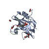 7rnsC 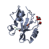 7rnuC 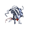 2iuiS S: Starting model for refinement C: citing same article ( |
|---|---|
| Similar structure data |


-Links
 PDBj
PDBj


- Assembly
| Deposited unit | 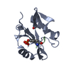
| ||||||||||||
|---|---|---|---|---|---|---|---|---|---|---|---|---|---|
| 1 |
| ||||||||||||
| Unit cell |
| ||||||||||||
| Components on special symmetry positions |
|
-Components
| #1: Protein | Mass: 13807.589 Da / Num. of mol.: 1 / Fragment: SH2 domain (UNP residues 665-774) Source method: isolated from a genetically manipulated source Source: (gene. exp.) Homo sapiens (human) / Gene: VAV2 / Production host:  | ||||||
|---|---|---|---|---|---|---|---|
| #2: Protein/peptide | Mass: 1063.975 Da / Num. of mol.: 1 / Fragment: UNP residues 52-60 / Source method: obtained synthetically / Source: (synth.) Homo sapiens (human) / References: UniProt: P68133 | ||||||
| #3: Chemical |   Mass: 62.068 Da / Num. of mol.: 2 / Source method: obtained synthetically / Formula: C2H6O2 Mass: 62.068 Da / Num. of mol.: 2 / Source method: obtained synthetically / Formula: C2H6O2#4: Water | ChemComp-HOH / |  Mass: 18.015 Da / Num. of mol.: 37 / Source method: isolated from a natural source / Formula: H2O Mass: 18.015 Da / Num. of mol.: 37 / Source method: isolated from a natural source / Formula: H2OHas ligand of interest | Y | Has protein modification | Y | |
-Experimental details
-Experiment
| Experiment | Method: X-RAY DIFFRACTION / Number of used crystals: 1 |
|---|
- Sample preparation
| Crystal | Density Matthews: 2.37 Å3/Da / Density % sol: 48.06 % |
|---|---|
| Crystal grow | Temperature: 292 K / Method: vapor diffusion, sitting drop / pH: 8.5 Details: 0.2 M sodium acetate trihydrate, 0.1 M Tris hydrochloride, pH 8.5, 30% PEG4000 |
-Data collection
| Diffraction | Mean temperature: 100 K / Serial crystal experiment: N |
|---|---|
| Diffraction source | Source: SEALED TUBE / Type: RIGAKU MICROMAX-003 / Wavelength: 1.54184 Å |
| Detector | Type: RIGAKU HyPix-6000HE / Detector: PIXEL / Date: Mar 28, 2019 |
| Radiation | Protocol: SINGLE WAVELENGTH / Monochromatic (M) / Laue (L): M / Scattering type: x-ray |
| Radiation wavelength | Wavelength: 1.54184 Å / Relative weight: 1 |
| Reflection | Resolution: 2.15→26.8 Å / Num. obs: 7715 / % possible obs: 96.1 % / Redundancy: 13.1 % / Biso Wilson estimate: 34.47 Å2 / CC1/2: 0.987 / Net I/σ(I): 10.3 |
| Reflection shell | Resolution: 2.15→2.23 Å / Num. unique obs: 5024 / CC1/2: 0.312 |
- Processing
| Software |
| ||||||||||||||||||||||||||||||||||||||||||
|---|---|---|---|---|---|---|---|---|---|---|---|---|---|---|---|---|---|---|---|---|---|---|---|---|---|---|---|---|---|---|---|---|---|---|---|---|---|---|---|---|---|---|---|
| Refinement | Method to determine structure: MOLECULAR REPLACEMENT Starting model: PDB entry 2IUI Resolution: 2.15→26.8 Å / SU ML: 0.3405 / Cross valid method: FREE R-VALUE / σ(F): 1.33 / Phase error: 35.8157 Stereochemistry target values: GeoStd + Monomer Library + CDL v1.2
| ||||||||||||||||||||||||||||||||||||||||||
| Solvent computation | Shrinkage radii: 0.9 Å / VDW probe radii: 1.11 Å / Solvent model: FLAT BULK SOLVENT MODEL | ||||||||||||||||||||||||||||||||||||||||||
| Displacement parameters | Biso mean: 41.47 Å2 | ||||||||||||||||||||||||||||||||||||||||||
| Refinement step | Cycle: LAST / Resolution: 2.15→26.8 Å
| ||||||||||||||||||||||||||||||||||||||||||
| Refine LS restraints |
| ||||||||||||||||||||||||||||||||||||||||||
| LS refinement shell |
|
