+ データを開く
- 基本情報
基本情報
| 登録情報 | データベース: PDB / ID: 7q5p | |||||||||
|---|---|---|---|---|---|---|---|---|---|---|
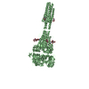
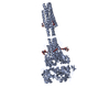
| タイトル | Structure of VgrG1 from Pseudomonas protegens. | |||||||||
 要素 要素 | Type VI secretion protein VgrG | |||||||||
 キーワード キーワード | TOXIN / bacterial type VI secretion system / VgrG | |||||||||
| 機能・相同性 | Bacteriophage T4 gp5 C-terminal trimerisation domain / Type VI secretion system, RhsGE-associated Vgr family subset / Type VI secretion system, RhsGE-associated Vgr protein / Phage tail baseplate hub (GPD) / Gp5/Type VI secretion system Vgr protein, OB-fold domain / Type VI secretion system/phage-baseplate injector OB domain / Vgr protein, OB-fold domain superfamily / : / Type VI secretion protein VgrG 機能・相同性情報 機能・相同性情報 | |||||||||
| 生物種 |  Pseudomonas fluorescens (蛍光菌) Pseudomonas fluorescens (蛍光菌) | |||||||||
| 手法 | 電子顕微鏡法 / 単粒子再構成法 / クライオ電子顕微鏡法 / 解像度: 3.3 Å | |||||||||
 データ登録者 データ登録者 | Guenther, P. / Quentin, D. / Ahmad, S. / Sachar, K. / Gatsogiannis, C. / Whitney, J.C. / Raunser, S. | |||||||||
| 資金援助 | 2件
| |||||||||
 引用 引用 |  ジャーナル: PLoS Pathog / 年: 2022 ジャーナル: PLoS Pathog / 年: 2022タイトル: Structure of a bacterial Rhs effector exported by the type VI secretion system. 著者: Patrick Günther / Dennis Quentin / Shehryar Ahmad / Kartik Sachar / Christos Gatsogiannis / John C Whitney / Stefan Raunser /   要旨: The type VI secretion system (T6SS) is a widespread protein export apparatus found in Gram-negative bacteria. The majority of T6SSs deliver toxic effector proteins into competitor bacteria. Yet, the ...The type VI secretion system (T6SS) is a widespread protein export apparatus found in Gram-negative bacteria. The majority of T6SSs deliver toxic effector proteins into competitor bacteria. Yet, the structure, function, and activation of many of these effectors remains poorly understood. Here, we present the structures of the T6SS effector RhsA from Pseudomonas protegens and its cognate T6SS spike protein, VgrG1, at 3.3 Å resolution. The structures reveal that the rearrangement hotspot (Rhs) repeats of RhsA assemble into a closed anticlockwise β-barrel spiral similar to that found in bacterial insecticidal Tc toxins and in metazoan teneurin proteins. We find that the C-terminal toxin domain of RhsA is autoproteolytically cleaved but remains inside the Rhs 'cocoon' where, with the exception of three ordered structural elements, most of the toxin is disordered. The N-terminal 'plug' domain is unique to T6SS Rhs proteins and resembles a champagne cork that seals the Rhs cocoon at one end while also mediating interactions with VgrG1. Interestingly, this domain is also autoproteolytically cleaved inside the cocoon but remains associated with it. We propose that mechanical force is required to remove the cleaved part of the plug, resulting in the release of the toxin domain as it is delivered into a susceptible bacterial cell by the T6SS. | |||||||||
| 履歴 |
|
- 構造の表示
構造の表示
| ムービー |
 ムービービューア ムービービューア |
|---|---|
| 構造ビューア | 分子:  Molmil Molmil Jmol/JSmol Jmol/JSmol |
- ダウンロードとリンク
- ダウンロード
| PDBx/mmCIF形式 |  7q5p.cif.gz 7q5p.cif.gz | 670.2 KB | 表示 |  PDBx/mmCIF形式 PDBx/mmCIF形式 |
|---|---|---|---|---|
| PDB形式 |  pdb7q5p.ent.gz pdb7q5p.ent.gz | 表示 |  PDB形式 PDB形式 | |
| PDBx/mmJSON形式 |  7q5p.json.gz 7q5p.json.gz | ツリー表示 |  PDBx/mmJSON形式 PDBx/mmJSON形式 | |
| その他 |  その他のダウンロード その他のダウンロード |
-検証レポート
| 文書・要旨 |  7q5p_validation.pdf.gz 7q5p_validation.pdf.gz | 874.5 KB | 表示 |  wwPDB検証レポート wwPDB検証レポート |
|---|---|---|---|---|
| 文書・詳細版 |  7q5p_full_validation.pdf.gz 7q5p_full_validation.pdf.gz | 892.5 KB | 表示 | |
| XML形式データ |  7q5p_validation.xml.gz 7q5p_validation.xml.gz | 68.1 KB | 表示 | |
| CIF形式データ |  7q5p_validation.cif.gz 7q5p_validation.cif.gz | 100.1 KB | 表示 | |
| アーカイブディレクトリ |  https://data.pdbj.org/pub/pdb/validation_reports/q5/7q5p https://data.pdbj.org/pub/pdb/validation_reports/q5/7q5p ftp://data.pdbj.org/pub/pdb/validation_reports/q5/7q5p ftp://data.pdbj.org/pub/pdb/validation_reports/q5/7q5p | HTTPS FTP |
-関連構造データ
- リンク
リンク
- 集合体
集合体
| 登録構造単位 | 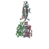
|
|---|---|
| 1 |
|
- 要素
要素
| #1: タンパク質 | 分子量: 72080.133 Da / 分子数: 3 / 由来タイプ: 組換発現 由来: (組換発現)  Pseudomonas fluorescens (strain ATCC BAA-477 / NRRL B-23932 / Pf-5) (バクテリア) Pseudomonas fluorescens (strain ATCC BAA-477 / NRRL B-23932 / Pf-5) (バクテリア)株: ATCC BAA-477 / NRRL B-23932 / Pf-5 / 遺伝子: vgrG, PFL_6094 / 発現宿主:  |
|---|
-実験情報
-実験
| 実験 | 手法: 電子顕微鏡法 |
|---|---|
| EM実験 | 試料の集合状態: PARTICLE / 3次元再構成法: 単粒子再構成法 |
- 試料調製
試料調製
| 構成要素 | 名称: Structure of the VgrG1 trimer from Pseudomonas protegens. タイプ: ORGANELLE OR CELLULAR COMPONENT / Entity ID: all / 由来: RECOMBINANT |
|---|---|
| 分子量 | 値: 0.215994 MDa / 実験値: NO |
| 由来(天然) | 生物種:  Pseudomonas protegens Pf-5 (バクテリア) Pseudomonas protegens Pf-5 (バクテリア) |
| 由来(組換発現) | 生物種:  |
| 緩衝液 | pH: 8 |
| 試料 | 濃度: 1 mg/ml / 包埋: NO / シャドウイング: NO / 染色: NO / 凍結: YES / 詳細: The sample was monodisperse. |
| 試料支持 | グリッドの材料: COPPER / グリッドのタイプ: Quantifoil R2/1 |
| 急速凍結 | 装置: FEI VITROBOT MARK IV / 凍結剤: ETHANE / 湿度: 100 % / 凍結前の試料温度: 281 K |
- 電子顕微鏡撮影
| 実験機器 |  モデル: Titan Krios / 画像提供: FEI Company |
|---|---|
| 顕微鏡 | モデル: FEI TITAN KRIOS |
| 電子銃 | 電子線源:  FIELD EMISSION GUN / 加速電圧: 300 kV / 照射モード: FLOOD BEAM FIELD EMISSION GUN / 加速電圧: 300 kV / 照射モード: FLOOD BEAM |
| 電子レンズ | モード: BRIGHT FIELD / 倍率(公称値): 59000 X / Cs: 0.01 mm / C2レンズ絞り径: 100 µm / アライメント法: COMA FREE |
| 試料ホルダ | 凍結剤: NITROGEN 試料ホルダーモデル: FEI TITAN KRIOS AUTOGRID HOLDER 最高温度: 70 K / 最低温度: 70 K |
| 撮影 | 平均露光時間: 1.5 sec. / 電子線照射量: 90 e/Å2 / 検出モード: INTEGRATING フィルム・検出器のモデル: FEI FALCON III (4k x 4k) 撮影したグリッド数: 1 / 実像数: 1250 |
| 電子光学装置 | エネルギーフィルター名称: GIF Bioquantum / エネルギーフィルタースリット幅: 20 eV / 球面収差補正装置: Cs corrector |
| 画像スキャン | 横: 4096 / 縦: 4096 |
- 解析
解析
| ソフトウェア | 名称: PHENIX / バージョン: dev_4338: / 分類: 精密化 | ||||||||||||||||||||||||||||||||||||
|---|---|---|---|---|---|---|---|---|---|---|---|---|---|---|---|---|---|---|---|---|---|---|---|---|---|---|---|---|---|---|---|---|---|---|---|---|---|
| EMソフトウェア |
| ||||||||||||||||||||||||||||||||||||
| CTF補正 | タイプ: PHASE FLIPPING AND AMPLITUDE CORRECTION | ||||||||||||||||||||||||||||||||||||
| 対称性 | 点対称性: C3 (3回回転対称) | ||||||||||||||||||||||||||||||||||||
| 3次元再構成 | 解像度: 3.3 Å / 解像度の算出法: FSC 0.143 CUT-OFF / 粒子像の数: 423980 / アルゴリズム: FOURIER SPACE / クラス平均像の数: 396 / 対称性のタイプ: POINT | ||||||||||||||||||||||||||||||||||||
| 原子モデル構築 | プロトコル: RIGID BODY FIT / 空間: REAL | ||||||||||||||||||||||||||||||||||||
| 原子モデル構築 | PDB-ID: 6H3N PDB chain-ID: A / Accession code: 6H3N / Pdb chain residue range: 1-643 / Source name: PDB / タイプ: experimental model |
 ムービー
ムービー コントローラー





 PDBj
PDBj
