 Movie
Movie Controller

[English] 日本語
 Yorodumi
- PDB-3maw: Structure of the Newcastle disease virus F protein in the post-fu... -
+ Open data

- Basic information
| Entry | Database: PDB / ID: 3maw | |||||||||
|---|---|---|---|---|---|---|---|---|---|---|
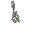
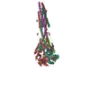
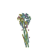
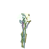
| Title | Structure of the Newcastle disease virus F protein in the post-fusion conformation | |||||||||
 Components Components | Fusion glycoprotein F0 | |||||||||
 Keywords Keywords | VIRAL PROTEIN / Fusion protein / class I / paramyxovirus / glycoprotein | |||||||||
| Function / homology |  Function and homology information Function and homology informationfusion of virus membrane with host plasma membrane / viral envelope / symbiont entry into host cell / host cell plasma membrane / virion membrane Similarity search - Function | |||||||||
| Biological species |  Newcastle disease virus Newcastle disease virus | |||||||||
| Method |  X-RAY DIFFRACTION / SYNCHROTRON / MOLECULAR REPLACEMENT / Resolution: 3.5 Å X-RAY DIFFRACTION / SYNCHROTRON / MOLECULAR REPLACEMENT / Resolution: 3.5 Å | |||||||||
 Authors Authors | Jardetzky, T.S. / Wen, X. | |||||||||
 Citation Citation | Journal: Virology / Year: 2010 Title: Structure of the Newcastle disease virus F protein in the post-fusion conformation. Authors: Swanson, K. / Wen, X. / Leser, G.P. / Paterson, R.G. / Lamb, R.A. / Jardetzky, T.S. | |||||||||
| History |
|
- Structure visualization
| Structure viewer | Molecule: MolmilJmol/JSmol |
|---|
- Downloads & links
-Download
| PDBx/mmCIF format | 3maw.cif.gz | 172 KB | Display | PDBx/mmCIF format |
|---|---|---|---|---|
| PDB format | pdb3maw.ent.gz | 133.3 KB | Display | PDB format |
| PDBx/mmJSON format | 3maw.json.gz | Tree view | PDBx/mmJSON format | |
| Others |  Other downloads Other downloads |
-Validation report
| Arichive directory | https://data.pdbj.org/pub/pdb/validation_reports/ma/3mawftp://data.pdbj.org/pub/pdb/validation_reports/ma/3maw | HTTPS FTP |
|---|
-Related structure data
| Similar structure data |
|---|

-Links
 PDBj
PDBj


- Assembly
| Deposited unit | 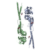
| ||||||||||||||||||
|---|---|---|---|---|---|---|---|---|---|---|---|---|---|---|---|---|---|---|---|
| 1 | 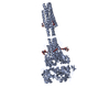
| ||||||||||||||||||
| 2 | 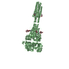
| ||||||||||||||||||
| Unit cell |
| ||||||||||||||||||
| Noncrystallographic symmetry (NCS) | NCS domain:
NCS domain segments:
|
-Components
| #1: Protein | Mass: 51164.852 Da / Num. of mol.: 2 / Fragment: UNP RESIDUE 32-499 Source method: isolated from a genetically manipulated source Source: (gene. exp.) Newcastle disease virus / Strain: Chicken/Australia-Victoria/32 / Gene: F / References: UniProt: P12572#2: Polysaccharide | 2-acetamido-2-deoxy-beta-D-glucopyranose-(1-4)-2-acetamido-2-deoxy-beta-D-glucopyranose Source method: isolated from a genetically manipulated source #3: Sugar |   Type: D-saccharide, alpha linking / Mass: 221.208 Da / Num. of mol.: 2 Type: D-saccharide, alpha linking / Mass: 221.208 Da / Num. of mol.: 2Source method: isolated from a genetically manipulated source Formula: C8H15NO6 Has protein modification | Y | Sequence details | H66L, I162N CONFLICT IN UNP ENTRY P12572 | |
|---|
-Experimental details
-Experiment
| Experiment | Method: X-RAY DIFFRACTION / Number of used crystals: 1 |
|---|
- Sample preparation
| Crystal | Density Matthews: 3.01 Å3/Da / Density % sol: 59.1 % |
|---|---|
| Crystal grow | Temperature: 277 K / Method: vapor diffusion, hanging drop / pH: 4.5 Details: 10% (wt/vol) polyethylene glycol 1000, 1.26 M ammonia sulfate, 0.1 M sodium acetate, pH 4.5, VAPOR DIFFUSION, HANGING DROP, temperature 277K |
-Data collection
| Diffraction | Mean temperature: 100 K |
|---|---|
| Diffraction source | Source: SYNCHROTRON / Site: APS  / Beamline: 5ID-B / Wavelength: 0.99998 Å / Beamline: 5ID-B / Wavelength: 0.99998 Å |
| Detector | Type: MARMOSAIC 225 mm CCD / Detector: CCD / Date: Jun 18, 2007 |
| Radiation | Monochromator: Si (111) / Protocol: SINGLE WAVELENGTH / Monochromatic (M) / Laue (L): M / Scattering type: x-ray |
| Radiation wavelength | Wavelength: 0.99998 Å / Relative weight: 1 |
| Reflection | Resolution: 3.5→50 Å / Num. obs: 14966 / % possible obs: 99.9 % / Observed criterion σ(F): 0 / Observed criterion σ(I): 0 / Redundancy: 6.2 % |
| Reflection shell | Resolution: 3.5→3.63 Å / % possible all: 100 |
- Processing
| Software |
| ||||||||||||||||||||||||||||||||||||||||||||||||||||||||
|---|---|---|---|---|---|---|---|---|---|---|---|---|---|---|---|---|---|---|---|---|---|---|---|---|---|---|---|---|---|---|---|---|---|---|---|---|---|---|---|---|---|---|---|---|---|---|---|---|---|---|---|---|---|---|---|---|---|
| Refinement | Method to determine structure: MOLECULAR REPLACEMENT / Resolution: 3.5→48.65 Å / SU ML: 0.64 / σ(F): 2 / Stereochemistry target values: ML
| ||||||||||||||||||||||||||||||||||||||||||||||||||||||||
| Solvent computation | Shrinkage radii: 0.9 Å / VDW probe radii: 1.11 Å / Solvent model: FLAT BULK SOLVENT MODEL / Bsol: 70.158 Å2 / ksol: 0.3 e/Å3 | ||||||||||||||||||||||||||||||||||||||||||||||||||||||||
| Refinement step | Cycle: LAST / Resolution: 3.5→48.65 Å
| ||||||||||||||||||||||||||||||||||||||||||||||||||||||||
| Refine LS restraints |
| ||||||||||||||||||||||||||||||||||||||||||||||||||||||||
| Refine LS restraints NCS |
| ||||||||||||||||||||||||||||||||||||||||||||||||||||||||
| LS refinement shell |
|
