 Movie
Movie Controller

[English] 日本語
 Yorodumi
- EMDB-0136: Electron cryo-microscopy of VgrG1 in the Type VI secretion VgrG1-... -
+ Open data

- Basic information
| Entry | Database: EMDB / ID: EMD-0136 | |||||||||
|---|---|---|---|---|---|---|---|---|---|---|
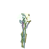
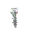
| Title | Electron cryo-microscopy of VgrG1 in the Type VI secretion VgrG1-Tse6-EF-Tu complex embedded in lipid nanodiscs | |||||||||
 Map data Map data | ||||||||||
 Sample Sample |
| |||||||||
 Keywords Keywords | Bacterial Type VI effector complex / Tse6-loaded VgrG1 complex / TOXIN | |||||||||
| Function / homology |  Function and homology information Function and homology informationprotein secretion by the type VI secretion system / type VI protein secretion system complex / extracellular region Similarity search - Function | |||||||||
| Biological species |  Pseudomonas aeruginosa PAO1 (bacteria) Pseudomonas aeruginosa PAO1 (bacteria) | |||||||||
| Method | single particle reconstruction / cryo EM / Resolution: 3.25 Å | |||||||||
 Authors Authors | Quentin D / Raunser S | |||||||||
| Funding support |  Germany, 1 items Germany, 1 items
| |||||||||
 Citation Citation | Journal: Nat Microbiol / Year: 2018 Title: Mechanism of loading and translocation of type VI secretion system effector Tse6. Authors: Dennis Quentin / Shehryar Ahmad / Premy Shanthamoorthy / Joseph D Mougous / John C Whitney / Stefan Raunser /   Abstract: The type VI secretion system (T6SS) primarily functions to mediate antagonistic interactions between contacting bacterial cells, but also mediates interactions with eukaryotic hosts. This molecular ...The type VI secretion system (T6SS) primarily functions to mediate antagonistic interactions between contacting bacterial cells, but also mediates interactions with eukaryotic hosts. This molecular machine secretes antibacterial effector proteins by undergoing cycles of extension and contraction; however, how effectors are loaded into the T6SS and subsequently delivered to target bacteria remains poorly understood. Here, using electron cryomicroscopy, we analysed the structures of the Pseudomonas aeruginosa effector Tse6 loaded onto the T6SS spike protein VgrG1 in solution and embedded in lipid nanodiscs. In the absence of membranes, Tse6 stability requires the chaperone EagT6, two dimers of which interact with the hydrophobic transmembrane domains of Tse6. EagT6 is not directly involved in Tse6 delivery but is crucial for its loading onto VgrG1. VgrG1-loaded Tse6 spontaneously enters membranes and its toxin domain translocates across a lipid bilayer, indicating that effector delivery by the T6SS does not require puncturing of the target cell inner membrane by VgrG1. Eag chaperone family members from diverse Proteobacteria are often encoded adjacent to putative toxins with predicted transmembrane domains and we therefore anticipate that our findings will be generalizable to numerous T6SS-exported membrane-associated effectors. | |||||||||
| History |
|
- Structure visualization
| Movie |
Movie viewer |
|---|---|
| Structure viewer | EM map: SurfViewMolmilJmol/JSmol |
| Supplemental images |

- Downloads & links
-EMDB archive
| Map data | emd_0136.map.gz | 9.7 MB | EMDB map data format | |
|---|---|---|---|---|
| Header (meta data) | emd-0136-v30.xmlemd-0136.xml | 12.3 KB 12.3 KB | Display Display | EMDB header |
| Images | 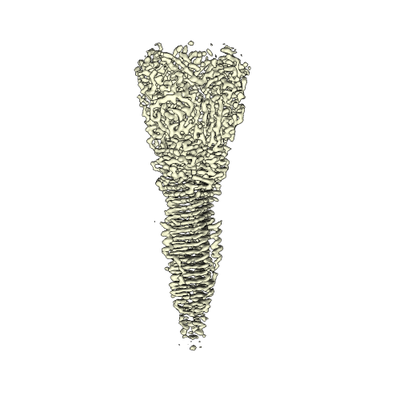 emd_0136.png emd_0136.png | 62.6 KB | ||
| Filedesc metadata | emd-0136.cif.gz | 5.9 KB | ||
| Archive directory |  http://ftp.pdbj.org/pub/emdb/structures/EMD-0136ftp://ftp.pdbj.org/pub/emdb/structures/EMD-0136 http://ftp.pdbj.org/pub/emdb/structures/EMD-0136ftp://ftp.pdbj.org/pub/emdb/structures/EMD-0136 | HTTPS FTP |
-Related structure data
| Related structure data |  6h3nMC  0135C  6h3lC M: atomic model generated by this map C: citing same article ( |
|---|---|
| Similar structure data |


-Links
| EMDB pages | EMDB (EBI/PDBe) / EMDataResource |
|---|
-Map
| File | Download / File: emd_0136.map.gz / Format: CCP4 / Size: 178 MB / Type: IMAGE STORED AS FLOATING POINT NUMBER (4 BYTES) | ||||||||||||||||||||||||||||||||||||||||||||||||||||||||||||
|---|---|---|---|---|---|---|---|---|---|---|---|---|---|---|---|---|---|---|---|---|---|---|---|---|---|---|---|---|---|---|---|---|---|---|---|---|---|---|---|---|---|---|---|---|---|---|---|---|---|---|---|---|---|---|---|---|---|---|---|---|---|
| Projections & slices | Image control
Images are generated by Spider. | ||||||||||||||||||||||||||||||||||||||||||||||||||||||||||||
| Voxel size | X=Y=Z: 1.1 Å | ||||||||||||||||||||||||||||||||||||||||||||||||||||||||||||
| Density |
| ||||||||||||||||||||||||||||||||||||||||||||||||||||||||||||
| Symmetry | Space group: 1 | ||||||||||||||||||||||||||||||||||||||||||||||||||||||||||||
| Details | EMDB XML:
CCP4 map header:
| ||||||||||||||||||||||||||||||||||||||||||||||||||||||||||||
 Z (Sec.)
 Y (Row.)
 X (Col.)


-Supplemental data
- Sample components
-Entire : The type VI secretion VgrG1-Tse6-EF-Tu complex embedded in lipid ...
| Entire | Name: The type VI secretion VgrG1-Tse6-EF-Tu complex embedded in lipid nanodiscs |
|---|---|
| Components |
|
-Supramolecule #1: The type VI secretion VgrG1-Tse6-EF-Tu complex embedded in lipid ...
| Supramolecule | Name: The type VI secretion VgrG1-Tse6-EF-Tu complex embedded in lipid nanodiscs type: complex / ID: 1 / Parent: 0 / Macromolecule list: all |
|---|---|
| Source (natural) | Organism: Pseudomonas aeruginosa PAO1 (bacteria) |
| Molecular weight | Theoretical: 350 KDa |
-Macromolecule #1: VgrG1
| Macromolecule | Name: VgrG1 / type: protein_or_peptide / ID: 1 / Number of copies: 3 / Enantiomer: LEVO |
|---|---|
| Source (natural) | Organism: Pseudomonas aeruginosa PAO1 (bacteria) |
| Molecular weight | Theoretical: 72.098375 KDa |
| Recombinant expression | Organism: |
| Sequence | String: MQLTRLVQVD CPLGPDVLLL QRMEGREELG RLFAYELHLV SENPNLPLEQ LLGKPMSLSL ELPGGSRRFF HGIVARCSQV AGHGQFAGY QATLRPWPWL LTRTSDCRIF QNQSVPEIIK QVFRNLGFSD FEDALTRPYR EWEYCVQYRE TSFDFISRLM E QEGIYYWF ...String: MQLTRLVQVD CPLGPDVLLL QRMEGREELG RLFAYELHLV SENPNLPLEQ LLGKPMSLSL ELPGGSRRFF HGIVARCSQV AGHGQFAGY QATLRPWPWL LTRTSDCRIF QNQSVPEIIK QVFRNLGFSD FEDALTRPYR EWEYCVQYRE TSFDFISRLM E QEGIYYWF RHEQKRHILV LSDAYGAHRS PGGYASVPYY PPTLGHRERD HFFDWQMARE VQPGSLTLND YDFQRPGARL EV RSNIARP HAAADYPLYD YPGEYVQSQD GEQYARNRIE AIQAQHERVR LRGVVRGIGA GHLFRLSGYP RDDQNREYLV VGA EYRVVQ ELYETGSGGA GSQFESELDC IDASQSFRLL PQTPVPVVRG PQTAVVVGPK GEEIWTDQYG RVKVHFHWDR HDQS NENSS CWIRVSQAWA GKNWGSMQIP RIGQEVIVSF LEGDPDRPII TGRVYNAEQT VPYELPANAT QSGMKSRSSK GGTPA NFNE IRMEDKKGAE QLYIHAERNQ DNLVENDASL SVGHDRNKSI GHDELARIGN NRTRAVKLND TLLVGGAKSD SVTGTY LIE AGAQIRLVCG KSVVEFNADG TINISGSAFN LYASGNGNID TGGRLDLNSG GASEVDAKGK GVQGTIDGQV QAMFPPP AK G UniProtKB: Type VI secretion system spike protein VgrG1a |
-Experimental details
-Structure determination
| Method | cryo EM |
|---|---|
 Processing Processing | single particle reconstruction |
| Aggregation state | particle |
-Sample preparation
| Concentration | 0.02 mg/mL | |||||||||
|---|---|---|---|---|---|---|---|---|---|---|
| Buffer | pH: 8 Component:
| |||||||||
| Grid | Model: Quantifoil R2/1 / Material: COPPER / Mesh: 400 / Support film - Material: CARBON / Support film - topology: CONTINUOUS / Support film - Film thickness: 2 / Pretreatment - Type: GLOW DISCHARGE | |||||||||
| Vitrification | Cryogen name: ETHANE / Chamber humidity: 95 % / Chamber temperature: 277 K / Instrument: FEI VITROBOT MARK III Details: 0.01 % Tween-20 was added to improve ice quality.. |
- Electron microscopy
| Microscope | FEI TITAN KRIOS |
|---|---|
| Specialist optics | Spherical aberration corrector: Cs corrected microscope |
| Image recording | Film or detector model: GATAN K2 SUMMIT (4k x 4k) / Detector mode: COUNTING / Number real images: 1873 / Average exposure time: 15.0 sec. / Average electron dose: 91.0 e/Å2 |
| Electron beam | Acceleration voltage: 300 kV / Electron source:  FIELD EMISSION GUN FIELD EMISSION GUN |
| Electron optics | Illumination mode: OTHER / Imaging mode: BRIGHT FIELD / Cs: 0.01 mm / Nominal defocus max: 3.2 µm / Nominal defocus min: 0.5 µm / Nominal magnification: 59000 |
| Sample stage | Specimen holder model: FEI TITAN KRIOS AUTOGRID HOLDER / Cooling holder cryogen: NITROGEN |
| Experimental equipment |  Model: Titan Krios / Image courtesy: FEI Company |
-Image processing
| Startup model | Type of model: EMDB MAP EMDB ID: |
|---|---|
| Final reconstruction | Applied symmetry - Point group: C3 (3 fold cyclic) / Resolution.type: BY AUTHOR / Resolution: 3.25 Å / Resolution method: FSC 0.143 CUT-OFF / Software - Name: SPHIRE / Software - details: meridien / Number images used: 72000 |
| Initial angle assignment | Type: MAXIMUM LIKELIHOOD |
| Final angle assignment | Type: MAXIMUM LIKELIHOOD |

