- PDB-1ztm: Structure of the Uncleaved Paramyxovirus (hPIV3) Fusion Protein -
+
Open data
ID or keywords:
Loading...
-
Basic information
Entry
Database: PDB / ID: 1ztm
Title
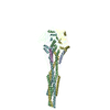
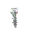
Structure of the Uncleaved Paramyxovirus (hPIV3) Fusion Protein
Components
Fusion glycoprotein
Keywords
VIRAL PROTEIN / fusion protein / 6-helix bundle / trimer / post-fusion
Function / homology
Function and homology information
fusion of virus membrane with host plasma membrane / viral envelope / symbiont entry into host cell / host cell plasma membrane / virion membrane Similarity search - Function
Helix Hairpins - #2480 / Head and neck region of the ectodomain of NDV fusion glycoprotein / Newcastle disease virus like domain / Head and neck region of the ectodomain of NDV fusion glycoprotein / YojJ-like / Precursor fusion glycoprotein F0, Paramyxoviridae / Fusion glycoprotein F0 / Helix Hairpins / Beta Barrel / Immunoglobulin-like ...Helix Hairpins - #2480 / Head and neck region of the ectodomain of NDV fusion glycoprotein / Newcastle disease virus like domain / Head and neck region of the ectodomain of NDV fusion glycoprotein / YojJ-like / Precursor fusion glycoprotein F0, Paramyxoviridae / Fusion glycoprotein F0 / Helix Hairpins / Beta Barrel / Immunoglobulin-like / Sandwich / Orthogonal Bundle / Mainly Beta / Mainly Alpha Similarity search - Domain/homology
In the structure databanks used in Yorodumi, some data are registered as the other names, "COVID-19 virus" and "2019-nCoV". Here are the details of the virus and the list of structure data.
Jan 31, 2019. EMDB accession codes are about to change! (news from PDBe EMDB page)
EMDB accession codes are about to change! (news from PDBe EMDB page)
The allocation of 4 digits for EMDB accession codes will soon come to an end. Whilst these codes will remain in use, new EMDB accession codes will include an additional digit and will expand incrementally as the available range of codes is exhausted. The current 4-digit format prefixed with “EMD-” (i.e. EMD-XXXX) will advance to a 5-digit format (i.e. EMD-XXXXX), and so on. It is currently estimated that the 4-digit codes will be depleted around Spring 2019, at which point the 5-digit format will come into force.
The EM Navigator/Yorodumi systems omit the EMD- prefix.
Related info.:Q: What is EMD? / ID/Accession-code notation in Yorodumi/EM Navigator
Yorodumi is a browser for structure data from EMDB, PDB, SASBDB, etc.
This page is also the successor to EM Navigator detail page, and also detail information page/front-end page for Omokage search.
The word "yorodu" (or yorozu) is an old Japanese word meaning "ten thousand". "mi" (miru) is to see.
Related info.:EMDB / PDB / SASBDB / Comparison of 3 databanks / Yorodumi Search / Aug 31, 2016. New EM Navigator & Yorodumi / Yorodumi Papers / Jmol/JSmol / Function and homology information / Changes in new EM Navigator and Yorodumi
 Movie
Movie Controller


 Open data
 Basic information
 Components
 Keywords
 Function and homology information
 Human parainfluenza virus 3
 X-RAY DIFFRACTION /
  Authors
 Citation
 Structure visualization
 Downloads & links
 Other downloads
 PDBj
PDBj

 Assembly
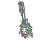
 Trichoplusia ni (cabbage looper) / References: UniProt: P06828
 Type: D-saccharide, beta linking / Mass: 221.208 Da / Num. of mol.: 2 / Source method: obtained synthetically / Formula: C8H15NO6
 Mass: 18.015 Da / Num. of mol.: 1 / Source method: isolated from a natural source / Formula: H2O
 Sample preparation
 Processing