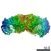
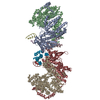
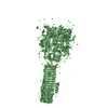
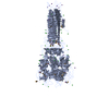
[English] 日本語
Yorodumi
- PDB-4uhv: The structure of VgrG1, the needle tip of the bacterial Type VI S... -

+
Open data


ID or keywords:

Loading...

-
Basic information

Entry
Database: PDB / ID: 4uhv
TitleThe structure of VgrG1, the needle tip of the bacterial Type VI Secretion System
ComponentsVGRG1, VALINE-GLYCINE REPEAT PROTEIN G1
KeywordsSTRUCTURAL PROTEIN / VGRG1 / VIRULENCE / TOXIN / EFFECTOR / PUNCTURING DEVICE / SPIKE / T6SS / P. AERUGINOSA
Function / homology
Function and homology information


type VI protein secretion system complex / protein secretion by the type VI secretion system / extracellular region
Similarity search - Function
Light-harvesting Protein - #110 / Pnp Oxidase; Chain A - #50 / Gp5 N-terminal domain / Baseplate protein-like domains / Light-harvesting Protein / Type VI secretion system, RhsGE-associated Vgr family subset / Phage tail baseplate hub (GPD) / Phage tail protein beta-alpha-beta fold / Type VI secretion system, RhsGE-associated Vgr protein / Gp5/Type VI secretion system Vgr protein, OB-fold domain ...Light-harvesting Protein - #110 / Pnp Oxidase; Chain A - #50 / Gp5 N-terminal domain / Baseplate protein-like domains / Light-harvesting Protein / Type VI secretion system, RhsGE-associated Vgr family subset / Phage tail baseplate hub (GPD) / Phage tail protein beta-alpha-beta fold / Type VI secretion system, RhsGE-associated Vgr protein / Gp5/Type VI secretion system Vgr protein, OB-fold domain / Type VI secretion system/phage-baseplate injector OB domain / Vgr protein, OB-fold domain superfamily / 3-Layer(bab) Sandwich / Pnp Oxidase; Chain A / Few Secondary Structures / Irregular / OB fold (Dihydrolipoamide Acetyltransferase, E2P) / Roll / Beta Barrel / Mainly Beta / Alpha Beta
Similarity search - Domain/homology
Type VI secretion system spike protein VgrG1a
Similarity search - Component
Biological speciesPSEUDOMONAS AERUGINOSA (bacteria)
MethodX-RAY DIFFRACTION / SYNCHROTRON / MOLECULAR REPLACEMENT / Resolution: 2 Å
AuthorsSpinola-Amilibia, M. / Davo-Siguero, I. / Ruiz, F.M. / Santillana, E. / Medrano, F.J. / Romero, A.
CitationJournal: Acta Crystallogr.,Sect.D / Year: 2016
Title: The Structure of Vgrg1 from Pseudomonas Aeruginosa, the Needle Tip of the Bacterial Type Vi Secretion System
Authors: Spinola-Amilibia, M. / Davo-Siguero, I. / Ruiz, F.M. / Santillana, E. / Medrano, F.J. / Romero, A.
History
DepositionMar 25, 2015Deposition site: PDBE / Processing site: PDBE
Revision 1.0Jan 27, 2016Provider: repository / Type: Initial release
Revision 1.1Mar 2, 2016Group: Database references
Revision 1.2Jan 10, 2024Group: Data collection / Database references ...Data collection / Database references / Derived calculations / Other / Refinement description
Category: chem_comp_atom / chem_comp_bond ...chem_comp_atom / chem_comp_bond / database_2 / pdbx_database_status / pdbx_initial_refinement_model / pdbx_struct_conn_angle / struct_conn / struct_site
Item: _database_2.pdbx_DOI / _database_2.pdbx_database_accession ..._database_2.pdbx_DOI / _database_2.pdbx_database_accession / _pdbx_database_status.status_code_sf / _pdbx_struct_conn_angle.ptnr1_auth_comp_id / _pdbx_struct_conn_angle.ptnr1_auth_seq_id / _pdbx_struct_conn_angle.ptnr1_label_asym_id / _pdbx_struct_conn_angle.ptnr1_label_atom_id / _pdbx_struct_conn_angle.ptnr1_label_comp_id / _pdbx_struct_conn_angle.ptnr1_label_seq_id / _pdbx_struct_conn_angle.ptnr2_auth_seq_id / _pdbx_struct_conn_angle.ptnr2_label_asym_id / _pdbx_struct_conn_angle.ptnr3_auth_comp_id / _pdbx_struct_conn_angle.ptnr3_auth_seq_id / _pdbx_struct_conn_angle.ptnr3_label_asym_id / _pdbx_struct_conn_angle.ptnr3_label_atom_id / _pdbx_struct_conn_angle.ptnr3_label_comp_id / _pdbx_struct_conn_angle.ptnr3_label_seq_id / _pdbx_struct_conn_angle.value / _struct_conn.conn_type_id / _struct_conn.id / _struct_conn.pdbx_dist_value / _struct_conn.pdbx_leaving_atom_flag / _struct_conn.ptnr1_auth_asym_id / _struct_conn.ptnr1_auth_comp_id / _struct_conn.ptnr1_auth_seq_id / _struct_conn.ptnr1_label_asym_id / _struct_conn.ptnr1_label_atom_id / _struct_conn.ptnr1_label_comp_id / _struct_conn.ptnr1_label_seq_id / _struct_conn.ptnr2_auth_asym_id / _struct_conn.ptnr2_auth_comp_id / _struct_conn.ptnr2_auth_seq_id / _struct_conn.ptnr2_label_asym_id / _struct_conn.ptnr2_label_atom_id / _struct_conn.ptnr2_label_comp_id / _struct_conn.ptnr2_label_seq_id / _struct_site.pdbx_auth_asym_id / _struct_site.pdbx_auth_comp_id / _struct_site.pdbx_auth_seq_id
Remark 650 HELIX DETERMINATION METHOD: AUTHOR PROVIDED.
Remark 700 SHEET DETERMINATION METHOD: AUTHOR PROVIDED.

-
Structure visualization

Structure viewerMolecule:
MolmilJmol/JSmol

Downloads & links

-
Assembly

Deposited unit
A: VGRG1, VALINE-GLYCINE REPEAT PROTEIN G1
B: VGRG1, VALINE-GLYCINE REPEAT PROTEIN G1
hetero molecules


Theoretical massNumber of molelcules
Total (without water)148,82953
Polymers147,1832
Non-polymers1,64651
Water11,602644
1
B: VGRG1, VALINE-GLYCINE REPEAT PROTEIN G1
hetero molecules

B: VGRG1, VALINE-GLYCINE REPEAT PROTEIN G1
hetero molecules

B: VGRG1, VALINE-GLYCINE REPEAT PROTEIN G1
hetero molecules


Theoretical massNumber of molelcules
Total (without water)223,91799
Polymers220,7753
Non-polymers3,14296
Water543
TypeNameSymmetry operationNumber
identity operation1_555x,y,z1
crystal symmetry operation2_755-y+2,x-y,z1
crystal symmetry operation3_775-x+y+2,-x+2,z1
Buried area53100 Å2
ΔGint-295.3 kcal/mol
Surface area102870 Å2
MethodPQS
2
A: VGRG1, VALINE-GLYCINE REPEAT PROTEIN G1
hetero molecules

A: VGRG1, VALINE-GLYCINE REPEAT PROTEIN G1
hetero molecules

A: VGRG1, VALINE-GLYCINE REPEAT PROTEIN G1
hetero molecules


Theoretical massNumber of molelcules
Total (without water)222,57160
Polymers220,7753
Non-polymers1,79657
Water543
TypeNameSymmetry operationNumber
identity operation1_555x,y,z1
crystal symmetry operation2_645-y+1,x-y-1,z1
crystal symmetry operation3_765-x+y+2,-x+1,z1
Buried area52980 Å2
ΔGint-282.6 kcal/mol
Surface area105410 Å2
MethodPQS
Unit cell
Length a, b, c (Å)78.620, 78.620, 430.320
Angle α, β, γ (deg.)90.00, 90.00, 120.00
Int Tables number173
Space group name H-MP63

-
Components

#1: Protein VGRG1, VALINE-GLYCINE REPEAT PROTEIN G1


Mass: 73591.602 Da / Num. of mol.: 2
Source method: isolated from a genetically manipulated source
Source: (gene. exp.) PSEUDOMONAS AERUGINOSA (bacteria) / Strain: PAO1 / Plasmid: PET29_VGRG1 / Production host: ESCHERICHIA COLI (E. coli) / Strain (production host): BL21(DE3) / Variant (production host): ROSETTA PLYSS / References: UniProt: Q9I741
#2: Chemical...
ChemComp-CL / CHLORIDE ION / Chloride


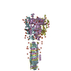
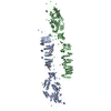
Mass: 35.453 Da / Num. of mol.: 38 / Source method: obtained synthetically / Formula: Cl
#3: Chemical
ChemComp-NA / SODIUM ION


Mass: 22.990 Da / Num. of mol.: 13 / Source method: obtained synthetically / Formula: Na
#4: Water ChemComp-HOH / water / Water


Mass: 18.015 Da / Num. of mol.: 644 / Source method: isolated from a natural source / Formula: H2O

-
Experimental details

-
Experiment

ExperimentMethod: X-RAY DIFFRACTION / Number of used crystals: 1

-
Sample preparation

CrystalDensity Matthews: 2.62 Å3/Da / Density % sol: 53.1 % / Description: TWINNED DATA, L-TEST OF 0.34
Crystal growpH: 7.2
Details: PROTEIN WAS CRYSTALLIZED FROM 0.1 M HEPES PH 7.2, 0.2 M LITHIUM SULPHATE AND 38 % MPD; THEN THE CRYSTAL WAS SOAKED IN 2 MM NEODYMIUM (III) SULFATE

-
Data collection

DiffractionMean temperature: 100 K
Diffraction sourceSource: SYNCHROTRON / Site: ALBA / Beamline: XALOC / Wavelength: 1.72785
DetectorType: DECTRIS PILATUS 6M / Detector: PIXEL / Date: Jul 24, 2014 / Details: KB MIRRORS
RadiationMonochromator: SI(111) / Protocol: SINGLE WAVELENGTH / Monochromatic (M) / Laue (L): M / Scattering type: x-ray
Radiation wavelengthWavelength: 1.72785 Å / Relative weight: 1
Reflection twin
Crystal-IDIDOperatorDomain-IDFraction
11H, K, L10.5
11K, H, -L20.5
ReflectionResolution: 1.88→86.06 Å / Num. obs: 111022 / % possible obs: 91.9 % / Observed criterion σ(I): -3 / Redundancy: 12.5 % / Rmerge(I) obs: 0.25 / Net I/σ(I): 6
Reflection shellResolution: 1.88→1.92 Å / Redundancy: 4 % / Rmerge(I) obs: 0.75 / Mean I/σ(I) obs: 1.1 / % possible all: 56.1

-
Processing

Software
NameVersionClassification
REFMAC5.8.0073refinement
XDSdata reduction
Aimlessdata scaling
PHASERphasing
RefinementMethod to determine structure: MOLECULAR REPLACEMENT
Starting model: PDB ENTRY 4MTK
Resolution: 2→71.73 Å / Cor.coef. Fo:Fc: 0.912 / Cor.coef. Fo:Fc free: 0.893 / SU B: 3.812 / SU ML: 0.113 / Cross valid method: THROUGHOUT / ESU R: 0.048 / ESU R Free: 0.038 / Stereochemistry target values: MAXIMUM LIKELIHOOD
Details: HYDROGENS HAVE BEEN ADDED IN THE RIDING POSITIONS. U VALUES REFINED INDIVIDUALLY
RfactorNum. reflection% reflectionSelection details
Rfree0.24845 4702 5 %RANDOM
Rwork0.22332 ---
obs0.22457 90119 93.98 %-
Solvent computationIon probe radii: 0.8 Å / Shrinkage radii: 0.8 Å / VDW probe radii: 1.2 Å / Solvent model: MASK
Displacement parametersBiso mean: 36.78 Å2
Baniso -1Baniso -2Baniso -3
1-5.92 Å20 Å20 Å2
2--5.92 Å20 Å2
3----11.84 Å2
Refinement stepCycle: LAST / Resolution: 2→71.73 Å
ProteinNucleic acidLigandSolventTotal
Num. atoms10189 0 51 644 10884
Refine LS restraints
Refine-IDTypeDev idealDev ideal targetNumber
X-RAY DIFFRACTIONr_bond_refined_d0.010.01910462
X-RAY DIFFRACTIONr_bond_other_d0.0020.029696
X-RAY DIFFRACTIONr_angle_refined_deg1.6621.95114185
X-RAY DIFFRACTIONr_angle_other_deg0.934322228
X-RAY DIFFRACTIONr_dihedral_angle_1_deg10.67351299
X-RAY DIFFRACTIONr_dihedral_angle_2_deg38.69223.43554
X-RAY DIFFRACTIONr_dihedral_angle_3_deg22.317151641
X-RAY DIFFRACTIONr_dihedral_angle_4_deg18.13115105
X-RAY DIFFRACTIONr_chiral_restr0.0980.21468
X-RAY DIFFRACTIONr_gen_planes_refined0.0090.02112254
X-RAY DIFFRACTIONr_gen_planes_other0.0010.022619
X-RAY DIFFRACTIONr_nbd_refined
X-RAY DIFFRACTIONr_nbd_other
X-RAY DIFFRACTIONr_nbtor_refined
X-RAY DIFFRACTIONr_nbtor_other
X-RAY DIFFRACTIONr_xyhbond_nbd_refined
X-RAY DIFFRACTIONr_xyhbond_nbd_other
X-RAY DIFFRACTIONr_metal_ion_refined
X-RAY DIFFRACTIONr_metal_ion_other
X-RAY DIFFRACTIONr_symmetry_vdw_refined
X-RAY DIFFRACTIONr_symmetry_vdw_other
X-RAY DIFFRACTIONr_symmetry_hbond_refined
X-RAY DIFFRACTIONr_symmetry_hbond_other
X-RAY DIFFRACTIONr_symmetry_metal_ion_refined
X-RAY DIFFRACTIONr_symmetry_metal_ion_other
X-RAY DIFFRACTIONr_mcbond_it1.8523.7355166
X-RAY DIFFRACTIONr_mcbond_other1.8513.7355165
X-RAY DIFFRACTIONr_mcangle_it2.7715.5996457
X-RAY DIFFRACTIONr_mcangle_other2.7715.5996458
X-RAY DIFFRACTIONr_scbond_it1.5263.7615296
X-RAY DIFFRACTIONr_scbond_other1.5243.765294
X-RAY DIFFRACTIONr_scangle_it
X-RAY DIFFRACTIONr_scangle_other2.2085.6337722
X-RAY DIFFRACTIONr_long_range_B_refined5.23229.99111876
X-RAY DIFFRACTIONr_long_range_B_other5.12729.98611798
X-RAY DIFFRACTIONr_rigid_bond_restr
X-RAY DIFFRACTIONr_sphericity_free
X-RAY DIFFRACTIONr_sphericity_bonded
LS refinement shellResolution: 2→2.052 Å / Total num. of bins used: 20
RfactorNum. reflection% reflection
Rfree0.314 313 -
Rwork0.249 6063 -
obs--85.86 %

+
About Yorodumi

-
News

-
Feb 9, 2022. New format data for meta-information of EMDB entries

New format data for meta-information of EMDB entries

  • Version 3 of the EMDB header file is now the official format.
  • The previous official version 1.9 will be removed from the archive.

Related info.:EMDB header

External links:wwPDB to switch to version 3 of the EMDB data model

-
Aug 12, 2020. Covid-19 info

Covid-19 info

URL: https://pdbj.org/emnavi/covid19.php

New page: Covid-19 featured information page in EM Navigator.

Related info.:Covid-19 info / Mar 5, 2020. Novel coronavirus structure data

+
Mar 5, 2020. Novel coronavirus structure data

Novel coronavirus structure data

Related info.:Yorodumi Speices / Aug 12, 2020. Covid-19 info

External links:COVID-19 featured content - PDBj / Molecule of the Month (242):Coronavirus Proteases

+
Jan 31, 2019. EMDB accession codes are about to change! (news from PDBe EMDB page)

EMDB accession codes are about to change! (news from PDBe EMDB page)

  • The allocation of 4 digits for EMDB accession codes will soon come to an end. Whilst these codes will remain in use, new EMDB accession codes will include an additional digit and will expand incrementally as the available range of codes is exhausted. The current 4-digit format prefixed with “EMD-” (i.e. EMD-XXXX) will advance to a 5-digit format (i.e. EMD-XXXXX), and so on. It is currently estimated that the 4-digit codes will be depleted around Spring 2019, at which point the 5-digit format will come into force.
  • The EM Navigator/Yorodumi systems omit the EMD- prefix.

Related info.:Q: What is EMD? / ID/Accession-code notation in Yorodumi/EM Navigator

External links:EMDB Accession Codes are Changing Soon! / Contact to PDBj

+
Jul 12, 2017. Major update of PDB

Major update of PDB

  • wwPDB released updated PDB data conforming to the new PDBx/mmCIF dictionary.
  • This is a major update changing the version number from 4 to 5, and with Remediation, in which all the entries are updated.
  • In this update, many items about electron microscopy experimental information are reorganized (e.g. em_software).
  • Now, EM Navigator and Yorodumi are based on the updated data.

External links:wwPDB Remediation / Enriched Model Files Conforming to OneDep Data Standards Now Available in the PDB FTP Archive

-
Yorodumi

Thousand views of thousand structures

  • Yorodumi is a browser for structure data from EMDB, PDB, SASBDB, etc.
  • This page is also the successor to EM Navigator detail page, and also detail information page/front-end page for Omokage search.
  • The word "yorodu" (or yorozu) is an old Japanese word meaning "ten thousand". "mi" (miru) is to see.

Related info.:EMDB / PDB / SASBDB / Comparison of 3 databanks / Yorodumi Search / Aug 31, 2016. New EM Navigator & Yorodumi / Yorodumi Papers / Jmol/JSmol / Function and homology information / Changes in new EM Navigator and Yorodumi

Read more